. 2014 Dec;273(3):746-58.
doi: 10.1148/radiol.14140033. Epub 2014 Jul 15.

Radiologic-pathologic analysis of contrast-enhanced and diffusion-weighted MR imaging in patients with HCC after TACE: diagnostic accuracy of 3D quantitative image analysis

Affiliations

Radiologic-pathologic analysis of contrast-enhanced and diffusion-weighted MR imaging in patients with HCC after TACE: diagnostic accuracy of 3D quantitative image analysis

Julius Chapiro et al. Radiology. 2014 Dec.

Abstract

Purpose: To evaluate the diagnostic performance of three-dimensional ( 3D three-dimensional ) quantitative enhancement-based and diffusion-weighted volumetric magnetic resonance (MR) imaging assessment of hepatocellular carcinoma ( HCC hepatocellular carcinoma ) lesions in determining the extent of pathologic tumor necrosis after transarterial chemoembolization ( TACE transarterial chemoembolization ).

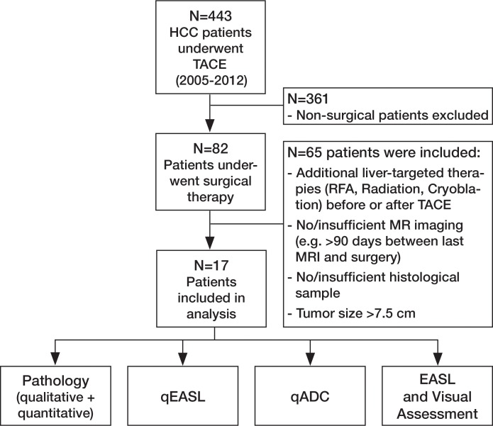
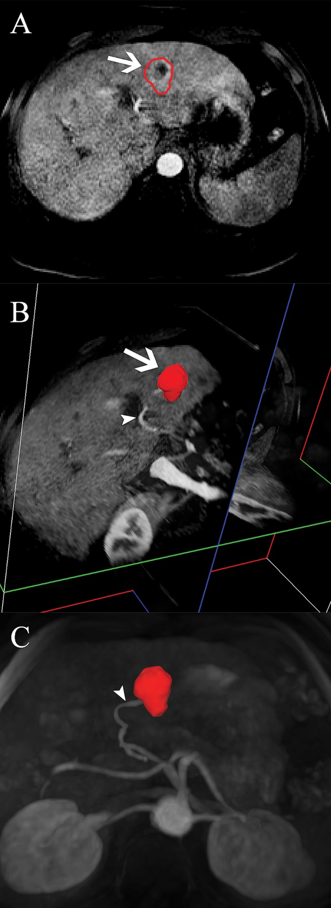
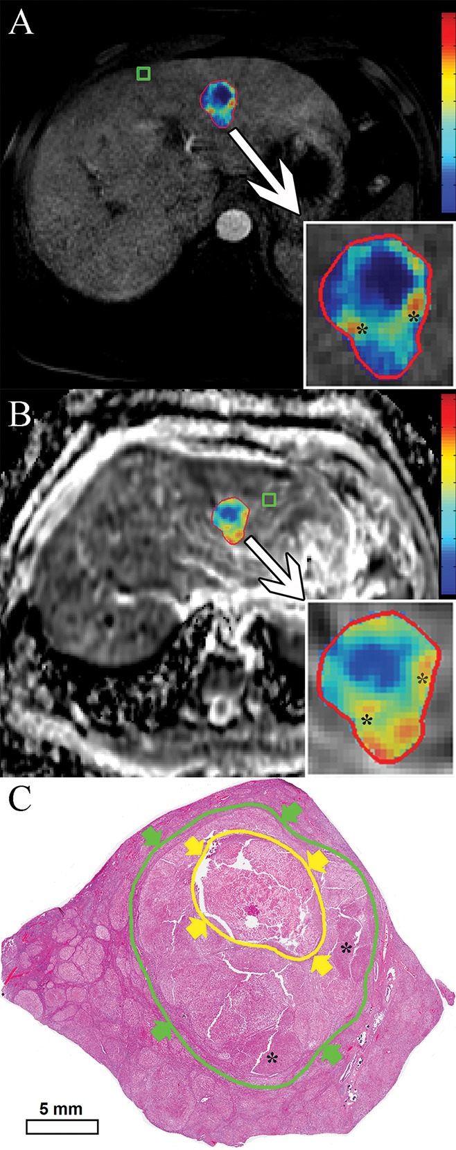
Materials and methods: This institutional review board-approved retrospective study included 17 patients with HCC hepatocellular carcinoma who underwent TACE transarterial chemoembolization before surgery. Semiautomatic 3D three-dimensional volumetric segmentation of target lesions was performed at the last MR examination before orthotopic liver transplantation or surgical resection. The amount of necrotic tumor tissue on contrast material-enhanced arterial phase MR images and the amount of diffusion-restricted tumor tissue on apparent diffusion coefficient ( ADC apparent diffusion coefficient ) maps were expressed as a percentage of the total tumor volume. Visual assessment of the extent of tumor necrosis and tumor response according to European Association for the Study of the Liver ( EASL European Association for the Study of the Liver ) criteria was performed. Pathologic tumor necrosis was quantified by using slide-by-slide segmentation. Correlation analysis was performed to evaluate the predictive values of the radiologic techniques.

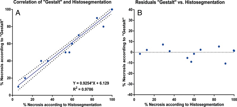
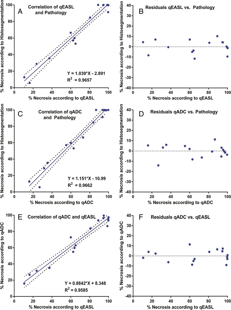
Results: At histopathologic examination, the mean percentage of tumor necrosis was 70% (range, 10%-100%). Both 3D three-dimensional quantitative techniques demonstrated a strong correlation with tumor necrosis at pathologic examination (R(2) = 0.9657 and R(2) = 0.9662 for quantitative EASL European Association for the Study of the Liver and quantitative ADC apparent diffusion coefficient , respectively) and a strong intermethod agreement (R(2) = 0.9585). Both methods showed a significantly lower discrepancy with pathologically measured necrosis (residual standard error [ RSE residual standard error ] = 6.38 and 6.33 for quantitative EASL European Association for the Study of the Liver and quantitative ADC apparent diffusion coefficient , respectively), when compared with non- 3D three-dimensional techniques ( RSE residual standard error = 12.18 for visual assessment).

Conclusion: This radiologic-pathologic correlation study demonstrates the diagnostic accuracy of 3D three-dimensional quantitative MR imaging techniques in identifying pathologically measured tumor necrosis in HCC hepatocellular carcinoma lesions treated with TACE transarterial chemoembolization .


Figures

Figure 1a:
Figure 1a:
(a) Flowchart of study design. Chart summarizes patient selection, exclusion criteria, and assessment techniques. Patients who received systemic or local-regional liver-targeted therapies other than TACE (n = 12) were excluded to avoid any non-TACE–related tumor response and/or necrosis. Patients who did not undergo follow-up MR imaging within 90 days before surgical treatment (n = 9) were excluded to avoid any potential discordance between imaging and tumor pathology owing to disease progression. Patients with severe motion artifacts on MR images affecting the target tumor regions were further excluded (n = 5). Only patients with well-defined index lesions smaller than 7.5 cm in largest diameter at baseline imaging were selected for further analysis. We excluded larger tumors (n = 24) because of the potential for bias in submission of representative pathologic sections of large tumors in this retrospective analysis. We also excluded patients in whom all pathologic slices of tumor were not available for review (n = 15). RFA = radiofrequency ablation. (b) Segmentation technique. A, Representative contrast-enhanced T1-weighted MR image demonstrates semiautomated tumor segmentation. Arrow = tumor rim. B, Three-dimensional model of upper abdomen shows volume rendering for segmented tumor. Arrow = tumor, arrowhead = branch of right hepatic artery that feeds tumor. C, Maximum intensity projection demonstrates contrast-enhanced blood vessels. Arrowhead = tumor-feeding artery.
Figure 1b:
Figure 1b:
(a) Flowchart of study design. Chart summarizes patient selection, exclusion criteria, and assessment techniques. Patients who received systemic or local-regional liver-targeted therapies other than TACE (n = 12) were excluded to avoid any non-TACE–related tumor response and/or necrosis. Patients who did not undergo follow-up MR imaging within 90 days before surgical treatment (n = 9) were excluded to avoid any potential discordance between imaging and tumor pathology owing to disease progression. Patients with severe motion artifacts on MR images affecting the target tumor regions were further excluded (n = 5). Only patients with well-defined index lesions smaller than 7.5 cm in largest diameter at baseline imaging were selected for further analysis. We excluded larger tumors (n = 24) because of the potential for bias in submission of representative pathologic sections of large tumors in this retrospective analysis. We also excluded patients in whom all pathologic slices of tumor were not available for review (n = 15). RFA = radiofrequency ablation. (b) Segmentation technique. A, Representative contrast-enhanced T1-weighted MR image demonstrates semiautomated tumor segmentation. Arrow = tumor rim. B, Three-dimensional model of upper abdomen shows volume rendering for segmented tumor. Arrow = tumor, arrowhead = branch of right hepatic artery that feeds tumor. C, Maximum intensity projection demonstrates contrast-enhanced blood vessels. Arrowhead = tumor-feeding artery.
Figure 2:
Figure 2:
Quantitative assessment techniques. A, qEASL color map of tumor (red represents maximum enhancement and blue represents no enhancement and/or necrotic tumor tissue, normalized by the ROI). B, qADC color map of same lesion (with color coding as in A). C, Digital scan (hematoxylin-eosin stain) obtained with histosegmentation technique. Yellow arrows and circle indicate necrosis, green arrows and circle indicate entire tumor. Corresponding highly viable areas of tumor are shown on all images (*).
Figure 3:
Figure 3:
Comparison of pathologic assessment techniques. A, Correlation and, B, residual plots of Gestalt-based assessment with histosegmentation according to a linear regression model.
Figure 4:
Figure 4:
Correlation of 3D quantitative techniques with pathologic examination. A, Correlation and, B, residual plots demonstrate results for qEASL. C, Correlation and, D, residual plots demonstrate results for qADC. E, Correlation and, F, residual plots show intermethod agreement between qEASL and qADC.
Figure 5:
Figure 5:
Correlation of subjective radiologic readings with pathologic examination. A, Correlation and, B, residual plots show results for reader 1. C, Correlation and, D, residual plots show results for reader 2. E, Correlation and, F, residual plots demonstrate agreement between readers.

References

    1. Bruix J, Sherman M, Llovet JM, et al. . Clinical management of hepatocellular carcinoma: conclusions of the Barcelona-2000 EASL conference. European Association for the Study of the Liver. J Hepatol 2001;35(3):421–430. - PubMed
    1. Lencioni R, Crocetti L. Local-regional treatment of hepatocellular carcinoma. Radiology 2012;262(1):43–58. - PubMed
    1. Forner A, Llovet JM, Bruix J. Hepatocellular carcinoma. Lancet 2012;379(9822):1245–1255. - PubMed
    1. European Association for the Study of the Liver; European Organisation for Research and Treatment of Cancer . EASL-EORTC clinical practice guidelines: management of hepatocellular carcinoma. J Hepatol 2012;56(4):908–943. - PubMed
    1. Chapiro J, Tacher V, Geschwind JF. Intraarterial therapies for primary liver cancer: state of the art. Expert Rev Anticancer Ther 2013;13(10):1157–1167. - PubMed

Publication types

LinkOut - more resources