Review
. 2014 Sep-Oct;49(5):413-25.
doi: 10.3109/10409238.2014.939608. Epub 2014 Jul 17.

The hypusine-containing translation factor eIF5A

Affiliations
Review

The hypusine-containing translation factor eIF5A

Thomas E Dever et al. Crit Rev Biochem Mol Biol. 2014 Sep-Oct.

Abstract

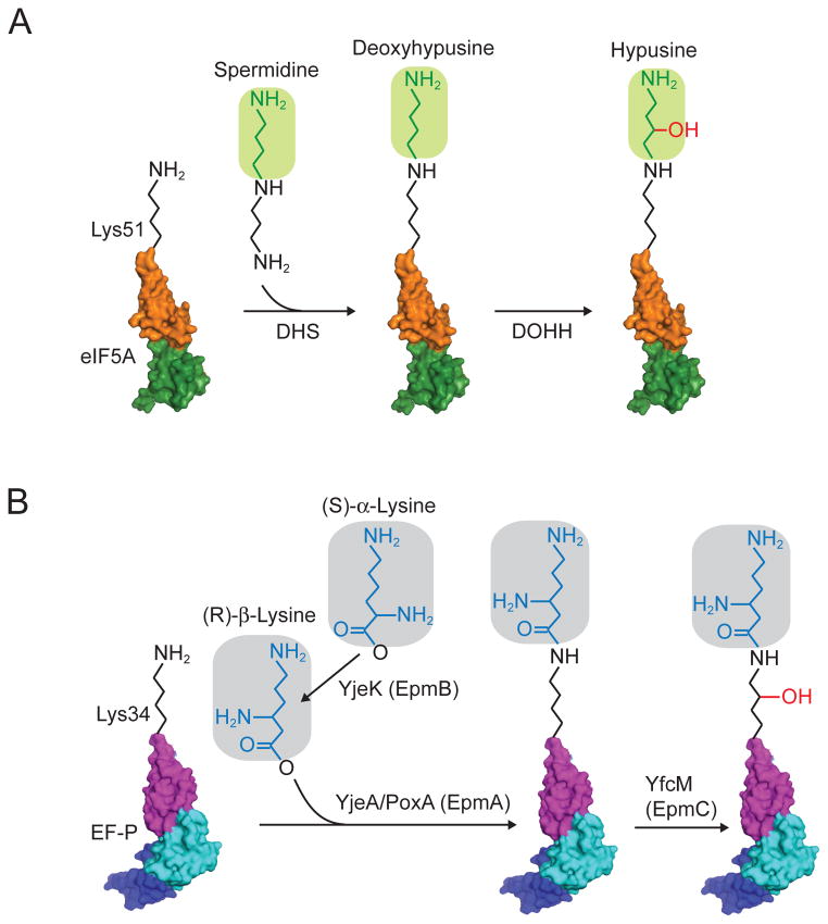
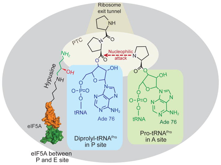
In addition to the small and large ribosomal subunits, aminoacyl-tRNAs, and an mRNA, cellular protein synthesis is dependent on translation factors. The eukaryotic translation initiation factor 5A (eIF5A) and its bacterial ortholog elongation factor P (EF-P) were initially characterized based on their ability to stimulate methionyl-puromycin (Met-Pmn) synthesis, a model assay for protein synthesis; however, the function of these factors in cellular protein synthesis has been difficult to resolve. Interestingly, a conserved lysine residue in eIF5A is post-translationally modified to hypusine and the corresponding lysine residue in EF-P from at least some bacteria is modified by the addition of a β-lysine moiety. In this review, we provide a summary of recent data that have identified a novel role for the translation factor eIF5A and its hypusine modification in the elongation phase of protein synthesis and more specifically in stimulating the production of proteins containing runs of consecutive proline residues.

Keywords: Deoxyhypusine hydroxylase; deoxyhypusine synthase; elongation factor; polyproline; translation elongation.


Conflict of interest statement

Declaration of interest

This work was supported by the Intramural Research Program of the National Institutes of Health, NICHD. The authors report no conflict of interest.

Figures

Figure 1
Figure 1. Schematic of the translation elongation cycle
(Top left) An EF-Tu(eukaryotic eEF1A)–GTP–aminoacyl(aa)-tRNA ternary complex (TC) binds aa-tRNA to the A site of the 70S (eukaryotic 80S) ribosome in a codon-dependent manner. (Top right) Following release of EF-Tu–GDP, the aa-tRNA is accommodated into the A site. Peptide bond formation (lower right) is followed by binding of EF-G (eEF2)–GTP (lower left), which promotes translocation of the deacylated tRNA and peptidyl tRNA into the canonical E and P sites, respectively. Following release of EF-G–GDP, the ribosome is ready for the next cycle of elongation. A color version of the figure is available online.
Figure 2
Figure 2. Sequence alignment of eIF5A and EF-P
Multiple sequence alignment of yeast (Saccharomyces cerevisiae, Sc) Hyp2 (Tif51A; eIF5A1) and Anb1 (Tif51B, eIF5A2), human (Hs) eIF5A1 and eIF5A2, fly (Drosophila melanogaster, Dm) eIF5A, archaeal (Methanococcus jannaschii, Mj, and Haloarcula marismortui, Hm) aIF5A, and bacterial (Escherichia coli, Ec, and Thermus thermophilus, Tt) EF-P was performed using Clustal X (v2.0). Residues in red are identical in all eIF5A and EF-P proteins; residues in blue are identical only in eIF5A and aIF5A; and residues in yellow are conserved in eIF5A and EF-P. Secondary structure elements are shown at the top (eIF5A) and bottom (EF-P) of the sequences, and colored based on the protein domain and scheme described in Figure 3. The lysine residue that is modified to hypusine in eIF5A and aIF5A and β-lysinylated in E. coli EF-P is colored black and denoted by the black dot above the alignment. A color version of the figure is available online.
Figure 3
Figure 3. Comparison of eIF5A and EF-P structures
Structures of Saccharomyces cerevisiae eIF5A (Left, PDB: 3ER0) and Thermus thermophilus EF-P (middle, PDB: 1UEB (Hanawa-Suetsugu et al., 2004)) are superimposed (right) using PyMOL software (PyMOL Molecular Graphics System, Version 1.5.0.4 Schrödinger, LLC). Colors are used to highlight the domains of eIF5A, N-terminal domain (orange) and C-terminal domain (green), and EF-P, domain I (magenta), domain II (cyan), and domain III (blue). The same color schemes are used in all figures. The modeled hypusine residue of eIF5A and corresponding Arg32 residue of EF-P are shown in stick presentation. A color version of the figure is available online.
Figure 4
Figure 4. Docking model of eIF5A–60S ribosome complex
(A) EF-P–70S ribosome complex (PDB: 3HUW and 3HUX, (Blaha, Stanley, and Steitz, 2009)). 50S subunit is colored gray and 30S subunit is colored light cyan, EF-P is colored as described in Figure 3, and tRNAfMet in P site is colored green. (Right) Magnified view of EF-P and P-site tRNAfMet structures. (B) Yeast eIF5A (PDB: 3ER0) was docked between the P and E sites of the yeast 60S ribosome (PDB: 3O30, (Ben-Shem et al., 2010)) by aligning with the structure of the EF-P–70S ribosome complex (panel A) and adjusting according to the results of hydroxyl radical cleavage data (Gutierrez et al., 2013). Positions of L1 stalk, P stalk and central protuberance (CP) on the 60S subunit are indicated; tRNAiMet is colored blue, and eIF5A is colored as described in Figure 3 with Lys51, the site of hypusine modification, colored black. (Right) Magnified view of docked eIF5A and P site tRNAiMet structures rotated 180°. A color version of the figure is available online.
Figure 5
Figure 5. Post-translational modification pathways for eIF5A and EF-P
(A) In eukaryotes and archaea, an n-butylamine moiety is transferred from spermidine to the ε-amino group of the specific lysine residue (Lys50 in human eIF5A and Lys51 in yeast) by the enzyme deoxyhypusine synthase (DHS). Next, deoxyhypusine hydroxylase (DOHH) adds a hydroxyl group to convert deoxyhypusine to hypusine. (B) In bacteria, β-lysinylation of EF-P occurs in three steps catalyzed by YjeK (EpmB), YjeA/PoxA (EpmA), and YfcM (EpmC). First, YjeK, a lysine aminomutase, converts lysine to β-lysine. Next, YjeA/PoxA catalyzes the addition of β-lysine onto the specific lysine residue (Lys34 on E. coli EF-P). Finally, YfcM hydroxylates the lysine residue. Domains of eIF5A and EF-P are colored as described in Figure 3. A color version of the figure is available online.
Figure 6
Figure 6. Model of eIF5A promoting polyproline peptide bond formation
Cartoon presentation of the large ribosomal subunit stalled with diprolyl-tRNAPro in the P site and Pro-tRNAPro in the A site. Binding of eIF5A between the P and E sites places the hypusine side chain near the diprolyl-tRNAPro in the peptidyl transferase center (PTC) of the ribosome where it facilitates peptide bond formation. Note that figure is not drawn to scale. A color version of the figure is available online.

References

    1. Aoki H, Xu J, Emili A, Chosay JG, Golshani A, Ganoza MC. Interactions of elongation factor EF-P with the Escherichia coli ribosome. FEBS J. 2008;275:671–81. - PubMed
    1. Bailly M, de Crecy-Lagard V. Predicting the pathway involved in post-translational modification of elongation factor P in a subset of bacterial species. Biol Direct. 2010;5:3. - PMC - PubMed
    1. Bartig D, Lemkemeier K, Frank J, Lottspeich F, Klink F. The archaebacterial hypusine-containing protein. Structural features suggest common ancestry with eukaryotic translation initiation factor 5A. Eur J Biochem. 1992;204:751–8. - PubMed
    1. Bartig D, Schumann H, Klink F. The unique posttranslational modification leading to deoxyhypusine or hypusine is a general feature of the archaebacterial kingdom. Syst Appl Microbiol. 1990;13:112–16.
    1. Ben-Shem A, Jenner L, Yusupova G, Yusupov M. Crystal structure of the eukaryotic ribosome. Science. 2010;330:1203–9. - PubMed

Publication types

MeSH terms

LinkOut - more resources