. 2014 Jul 17;511(7509):319-25.
doi: 10.1038/nature13535. Epub 2014 Jun 25.

The cancer glycocalyx mechanically primes integrin-mediated growth and survival

Affiliations

The cancer glycocalyx mechanically primes integrin-mediated growth and survival

Matthew J Paszek et al. Nature. .

Abstract

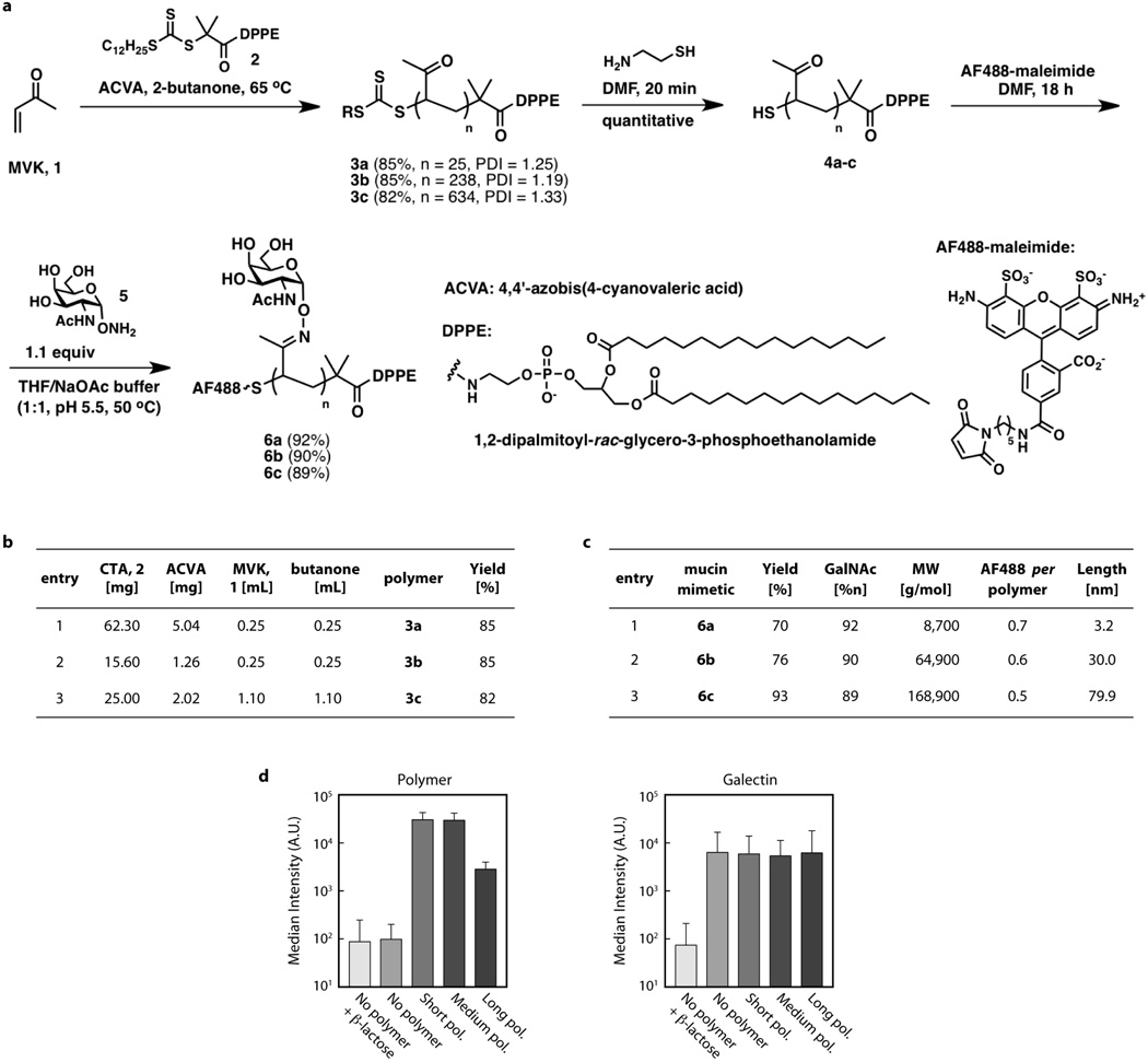
Malignancy is associated with altered expression of glycans and glycoproteins that contribute to the cellular glycocalyx. We constructed a glycoprotein expression signature, which revealed that metastatic tumours upregulate expression of bulky glycoproteins. A computational model predicted that these glycoproteins would influence transmembrane receptor spatial organization and function. We tested this prediction by investigating whether bulky glycoproteins in the glycocalyx promote a tumour phenotype in human cells by increasing integrin adhesion and signalling. Our data revealed that a bulky glycocalyx facilitates integrin clustering by funnelling active integrins into adhesions and altering integrin state by applying tension to matrix-bound integrins, independent of actomyosin contractility. Expression of large tumour-associated glycoproteins in non-transformed mammary cells promoted focal adhesion assembly and facilitated integrin-dependent growth factor signalling to support cell growth and survival. Clinical studies revealed that large glycoproteins are abundantly expressed on circulating tumour cells from patients with advanced disease. Thus, a bulky glycocalyx is a feature of tumour cells that could foster metastasis by mechanically enhancing cell-surface receptor function.


Figures

Extended Data Figure 1
Extended Data Figure 1. Large-scale gene expression analysis reveals increased expression of genes encoding bulky glycoproteins and glycan-modifying enzymes in primary tumours of patients with disseminated disease
a, Bioinformatics pipeline to estimate the extracellular bulkiness of a protein from its corresponding amino acid sequence. For each isoform sequence, the transmembrane and extramembrane domains were identified using a hidden Markov model (TMHMM). A combination of motif searches and neural network prediction then identified likely N- and O-glycosylation sites within each sequence. Isoform-level bulkiness estimates were generated by summing the number of predicted N- and O-glycosylation sites located within the extramembrane regions of the isoform. b, Heat map depicting the pairwise spearman correlation coefficients calculated by comparing all per-gene estimates of the total number of extra-membrane amino acids (AAoutside), N-glycosylation sites (Nglyc), O-glycosylation sites (Oglyc), and the overall bulkiness measure (total sites; for example, the sum of extra-membrane N- and O- glycosylation sites). Correlation coefficients relating the corresponding gene-wise measures are listed in the corresponding cells and depicted on a colour scale, where white corresponds to perfect correlation (rho = 1), and the dendrograms indicate the overall relationship between the parameters, estimated by Euclidean distance. High correlation coefficients indicate that gene-wise estimates of the compared parameters are similarly ranked (for example, genes with high values of X also tend to have high values of Y). The data indicate that the number of extracellular N-glycosylation sites and O-glycosylation sites identified within a gene are only weakly correlated, and neither dominates the total number of sites estimated per gene. c, Violin plots contrasting the distributions of gene-wise one-sided P values (y axis) quantifying evidence for transcriptional upregulation of glycosidases and glycosyltransferases, and subsets of glycosyltransferases (sialyltransferases and N-acetylgalactosaminyltransferases) with the full distribution. White dots and thick black lines indicate the median and interquartile range of the gene-wise P-value distribution among category members, and the width of the violin along the y axis indicates the density of the corresponding values. P values are derived from comparisons of expression levels in primary tumours of patients with or without distant metastases using a t-test. Indicated P values were estimated using a one-sided Kolmogorov–Smirnov test. d, Violin plots quantifying transcriptional upregulation of glycan-modifying enzymes in primary tumours of patients presenting with circulating tumour cells compared to tumours without detectable circulating tumour cells. e, Table of bulky glycoproteins and potential bulk-adding glycosyltransferases whose expression is upregulated in tumours that present with circulating tumour cells.
Extended Data Figure 2
Extended Data Figure 2. Computational model of the cell–ECM interface
Schematic of an integrated model that describes how the physical properties of the glycocalyx influence integrin–ECM interactions. The cell surface is modelled as a three-dimensional elastic plate; the ECM as a rigid substrate underneath the cell surface; and the glycocalyx as a repulsive potential between the plate and substrate. To compute stress–strain behaviour, the model is discretized using the three-dimensional lattice spring method, the cross-section of which is depicted above. Integrins are tethered to the cell surface and their distance-dependent binding to the ECM–substrate is calculated according to the Bell model. To calculate integrin-binding rate as a function of lateral distance from an adhesion cluster, an adhesion cluster is first constructed by assembling a 3 × 3 bond structure. The rates for additional integrin–ECM bonds then are computed at various distances from the cluster.
Extended Data Figure 3
Extended Data Figure 3. Synthesis and characterization of glycoprotein mimetics
a, Scheme for synthesis of lipid-terminated mucin mimetics labelled with Alexa Fluor 488 (AF488). b, Reagents and yields for the synthesis of polymers 3a–c. c, Characteristics of polymers 6a–c based on 1H NMR spectra. Glycoprotien mimetics were engineered to have minimal biochemical interactivity with cell surface lectins. d, Flow cytometry results quantifying incorporation of polymer on the surface of mammary epithelial cells (left) and binding with recombinant Alexa568-labelled galectin-3 with or without competitive inhibitor, β-lactose (right). Although a weak affinity between galectin-3 and the pendant N-acetylgalactosimes has previously been reported, the results suggest that incorporation of polymer does not significantly change the affinity of the cell surface for lectins.
Extended Data Figure 4
Extended Data Figure 4. MUC1 expression constructs
a, Schematic of MUC1 expression constructs. Full-length MUC1 consists of a large ectodomain with 42 mucin-type tandem repeats, a transmembrane domain, and short cytoplasmic tail. The tandem repeats and cytoplasmic tail are deleted in MUC1(ΔTR) and MUC1(ΔCT), respectively. For fluorescent protein fusions, mEmerald (GFP) and mEOS2 are fused to the C terminus of full-length MUC1 or MUC1(ΔCT). b, Schematic of MUC1 strain sensor and control constructs. Cysteine-free mTurqoiuse2 (CFP), Venus (YFP), or a FRET module consisting of the fluorescent proteins separated by an elastic linker (8 repeats of GPGGA) are inserted into the MUC1 ectodomain adjacent to the MUC1 tandem repeats. The mucin tandem repeats are deleted in ectodomain-truncated variants (ΔTR).
Extended Data Figure 5
Extended Data Figure 5. MUC1-mediated adhesion formation
a, Quantification of the average number of large adhesions, greater than 1 µm2, per area of cell in control epithelial cells (Control) and those ectopically expressing ectodomain-truncated MUC1 (+MUC1(ΔTR)), wild-type MUC1 (+MUC1), or cytoplasmic-tail-deleted MUC1 (+MUC1(ΔCT)). Results are the mean ± s.e.m. of three separate experiments. b, Fluorescence micrographs showing immuno-labelled MUC1 and fluorescently labelled fibronectin fibrils in control and MUC1-expressing epithelial cells. Soluble, labelled fibronectin in the growth media was deposited by cells at sites of cell–matrix adhesion. Binding of soluble fibronectin to MUC1 was not detected. Scale bar, 10 µm. c, Time lapse images of MUC1–YFP and vinculin–mCherry, showing the dynamics of adhesion assembly (Vinc.) and MUC1 patterning (MUC1). Scale bar, 1 µm. d, Rate of adhesion measured with single cell force spectroscopy of control (Cont.), α5 integrin-blocked (anti-α5), and MUC1-expressing cells (+MUC1) to fibronectin-coated surfaces and control cells to BSA-coated surfaces (BSA). Results are the mean ± s.e.m. of at least 15 cell measurements. Statistical significance is given by *P < 0.05; **P < 0.01; ***P < 0.001.
Extended Data Figure 6
Extended Data Figure 6. β3 integrin mobility in MUC1-expressing cells
a, Molecular diffusivity and adhesion enrichment measured with sptPALM in mouse embryonic fibroblasts (MEFs). Adhesion enrichment is reported as the ratio of the number of molecules detected inside focal adhesions per unit area to the number of molecules detected outside focal adhesions per unit area. b, Mean diffusion coefficients measured for freely diffusive β3 integrin tracks outside of adhesive contacts in control (Cont.) and MUC1-transfected (+MUC1) MEFs with and without Mn2+ to activate β3. c, Mean diffusion coefficients measured for confined β3 integrin tracks outside of adhesive contacts in MEFs with and without Mn2+. d, Mean radius of confinement measured for confined β3 integrin tracks outside of adhesive contacts in MEFs with and without Mn2+. e, Fraction of immobilized (Imm.), confined (Conf.), and freely diffusive (Free) β3 integrins inside of adhesive contacts in control and MUC1-transfected MEFs with and without Mn2+ treatment. f, From left to right, panels show GFP-tagged wild-type MUC1 (red) and positions of individual β3 integrins (green) in MEFs without Mn2+ treatment (left panel) and individual integrin trajectories recorded with sptPALM within MUC1-rich regions, outside MUC1-rich regions, and that cross MUC1 boundaries (scale bar, 2 µm). The ratio of integrins crossing out versus crossing in the MUC1 boundaries per cell is close to one (1.0 ± 0.1, n = 9 cells, 4,145 trajectories) showing that the flux of free diffusing integrins crossing in or out the mucin region is the same. g, From left to right, panels show integrin trajectories within an arbitrary region drawn in a MUC1-rich area (dashed white circles), outside of the circled region, and that cross the circled region (scale bar, 2 µm). The ratio of integrins crossing the MUC1-rich boundaries versus the fictive boundaries per cell is close to one (1.2 ± 0.2, n = 9 cells, 9,321 trajectories), showing that the MUC1–adhesive zone boundary does not affect the diffusive crossing of integrins. For all bar graphs, results are the mean ± s.e.m.
Extended Data Figure 7
Extended Data Figure 7. MUC1 strain gauge
a, Western blot of indicated construct expressed in HEK 293T cells and probed with anti-GFP family antibody, or full-length MUC1 construct expressed in HEK 293T cells and probed with an antibody against the MUC1 tandem repeats. b, Pseudocoloured images showing similar FRET efficiencies measured by the photobleaching FRET method for mammary epithelial cells (MECs) expressing low (Low) and high (High) levels of the sensor construct. Scale bar, 5 µm. c, Plot showing the level of CFP bleaching per CFP imaging cycle in MECs. d, Control images showing minimal intermolecular FRET in MECs expressing similar levels of bothMUC1 CFP and MUC1 YFP. e, Micrographs showing the emitted photons from CFP and their fluorescence lifetimes in MECs expressing ectodomain-truncated (MUC1(ΔTR) sensor) or full-length MUC1 strain sensors (MUC1 sensor). Shorter lifetimes are indicative of higher energy transfer between the CFP donor and YFP acceptor, and thus closer spatial proximity of the donor and acceptor (scale bar, 10 µm). f, Representative profile of CFP lifetimes and emitted photons of the full-length MUC1 sensor along the red line in panel e. Pixels 0 and 40 correspond to the base and tip of the arrow, respectively. A drop in fluorescence lifetime (Lifetime) is often observed before the drop in MUC1 molecular density (Photons) as an adhesive zone is approached.
Extended Data Figure 8
Extended Data Figure 8. Tension-dependent integrin activation and focal adhesion assembly in MUC1-expressing cells
a, Fluorescence micrographs of fibronectin-crosslinked α5 integrin in control and MUC1-expressing mammary epithelial cells (MECs) treated with solvent alone (DMSO), myosin-II inhibitor (blebbistatin; 50 µM), or Rho kinase inhibitor (Y-27632; 10 µM) for 1 h and detergent-extracted following crosslinking. Only fibronectin-bound integrins under mechanical tension are crosslinked and visualized following detergent extraction (scale bar, 15 µm). b, Fluorescence micrographs showing formation of myosin-independent adhesion complexes in MUC1-expressing MECs. Cells were pre-treated for 1 h and plated for 2 h in 50 µM blebbistatin (scale bar, 10 µm). c, Fluorescence micrographs of paxillin–mCherry and immuno-labelled activated FAK (pY397) in control and MUC1(ΔCT) expressing MECs plated on compliant fibronectin-conjugated hydrogels (E = 140 Pa; scale bar, 3 µm; ROI scale bar, 0.5 µm). d, Western blots showing phosphorylation of paxillin (pY118) in control and MUC1-expressing MECs on compliant substrates (E = 140 Pa) following overnight serum starvation and stimulation with EGF. MUC1-expressing cells treated with a pharmacological inhibitor of focal adhesion kinase (+FAKi) for 1 h before EGF stimulation did not exhibit robust paxillin phosphorylation.
Extended Data Figure 9
Extended Data Figure 9. Cell proliferation on soft ECM
a, Fluorescence micrographs showing DAPI-stained nuclei of control and MUC1(ΔCT)-expressing MECs after 24 h of plating on soft, fibronectin-conjugated hydrogels (E = 140 Pa; scale bar, 250 µm). The majority of cells plated as single cells, indicating that multi-cell colonies that formed at later time points were largely attributed to cell proliferation. b, Quantification of cell proliferation of MUC1(ΔCT)-expressing epithelial cells on soft hydrogels conjugated with bovine serum albumin (BSA) or fibronectin (Fn). Cells plated similarly on BSA– and Fn–hydrogels, but cell proliferation was significantly enhanced on Fn–hydrogels. Results are the mean ± s.e.m with statistical significance given by *P < 0.05; **P < 0.01; ***P < 0.001.
Extended Data Figure 10
Extended Data Figure 10. Hyaluronic acid production by tumour cells promotes cellular growth
a, Quantification of hyaluronic acid (HA) cell surface levels on control (10A-Cont.), transformed (10A-v-Src, 10A-HRAS) and malignant (MCF7, T47D) mammary epithelial cells (MECs). b, Fluorescence micrographs of HA and immuno-labelled paxillin on the v-Src transformed MECs (scale bars, 3 µm). c, Quantification of the number of v-Src-transformed MECs per colony 48 h after plating on soft polyacrylamide gels (fibronectin-conjugated) and treated with vehicle (DMSO), hyaluronic acid synthesis inhibitor 4-methylumbelliferone (+4MU; 0.3 µM), or competitive inhibitor HA oligonucleotides (+Oligo; 12-mer average oligonucleotide size; 100 mg ml−1). Results are the mean ± s.e.m with statistical significance is given by *P < 0.05; **P < 0.01; ***P < 0.001.
Figure 1
Figure 1. The cancer glycocalyx drives integrin clustering
a, Violin plots showing increased expression of genes encoding bulky transmembrane proteins in primary tumours of patients with distant metastases relative to those with local invasion. White dots and thick black lines indicate the median and interquartile range of the P value distribution of all transcripts within each class: all genes, all membrane proteins (Mem.), and bulky transmembrane proteins (Bulky). b, Computed relative rate of integrin–ECM ligand bond formation as a function of distance from a pre-existing adhesion cluster. c, Model of proposed glycocalyx-mediated integrin clustering. Shorter distances between integrin–ligand pairs result in faster kinetic rates of binding. d, Cartoon showing structure of glycoprotein mimetics with lipid insertion domain. e, Fluorescence micrographs of MEC adhesion complexes (vinculin–mCherry) and glycomimetics of the indicated length (scale bar, 3 µm). f, SAIM images of DiI-labelled ventral plasma membrane topography in MECs incorporated with glycomimetics (scale bar, 2.5 µm). g, Rate of integrin–substrate adhesion measured using single cell force spectroscopy in MECs with incorporated glycomimetics. h, Quantification of the total adhesion complex area per cell in MECs with incorporated glycomimetics. All results are the mean ± s.e.m. of three separate experiments. Statistical significance is given by *P < 0.05; **P < 0.01; ***P < 0.001.
Figure 2
Figure 2. The bulky cancer-associated glycoprotein MUC1 drives integrin clustering
a, Cartoon of MUC1 and quantification of MUC1 cell-surface levels on control (10A-Cont.), transformed (10A-v-Src, 10A-HRAS) and tumour (MCF7, T47D) cells. b, Topographical SAIM images of representative mCherry–CAAX-labelled ventral plasma membranes in control and MUC1–GFP-expressing (+MUC1) MECs (Scale bar, 5 µm; region of interest (ROI) scale bar, 2 µm). c, Quantification of mean plasma membrane height in control MECs and those ectopically expressing ectodomain-truncated MUC1–GFP (+MUC1(ΔTR)) and wild-type MUC1–GFP (+MUC1). Results are the mean ± s.e.m. of at least 15 cell measurements in duplicate experiments. d, Fluorescence micrographs of MUC1 (ΔTR) or wild-type MUC1 expressed in MECs and their focal adhesions labelled with vinculin–mCherry (scale bar, 3 µm; ROI scale bar, 1.5 µm). e, Quantification of the total adhesion complex area per cell in control non-malignant MECs (control) and those ectopically expressing MUC1 (ΔTR), wild-type MUC1, or cytoplasmic-tail-deleted MUC1 (+MUC1(ΔCT)). Results are the mean ± s.e.m. of three separate experiments. f, Left panel: trajectories of individual mEOS2-tagged MUC1 proteins recorded at 50 Hz using sptPALM (green) and focal adhesions visualized with paxillin–GFP (red) in MEFs (scale bar, 3 µm). Right panel: the ROI from the left panel with individual MUC1 tracks displayed in multiple colours (scale bar, 1 µm). Statistical significance is given by *P < 0.05; **P < 0.01; ***P < 0.001.
Figure 3
Figure 3. Bulky glycoproteins spatially regulate immobilization of activated integrins
a, Left panels: fluorescence micrographs displaying paxillin–GFP-labelled focal adhesions in control cells or MUC1-rich regions in MUC1–GFP-expressing MEFs, and positions of individual mEOS2-fused β3 integrins. Cells were treated without or with Mn2+ to activate integrins (scale bar, 3 µm). Right panels: magnified area of interest showing fluorescence micrographs of focal adhesions visualized with paxillin–GFP in control MEFs or MUC1 in MUC1–GFP-expressing MEFs, and individual β3 integrin trajectories recorded with sptPALM. Single molecule trajectories are colour-coded to indicate immobile and mobile (confined and freely diffusive) β3 integrins (scale bar, 1 µm). b, Distribution of β3 integrin diffusion coefficients recorded before or after Mn2+ treatment in control MEFs outside of adhesive contacts (left), MUC1-transfected MEFs inside MUC1-rich areas (middle), and MUC1-transfected MEFs outside MUC1-rich areas, including adhesive contacts (right). c, Fraction of immobilized, confined and freely diffusive β3 integrins outside of adhesive contacts in control MEFs (Ctrl) and MUC1-transfected cells (MUC1) before and after Mn2+ treatment. Results are the mean ± s.e.m. Statistical significance is given by *P < 0.05; **P < 0.01; ***P < 0.001. d, Fluorescence micrograph of MUC1–GFP and an illustrative single integrin trajectory in MEFs treated with Mn2+ (scale bar, 1 µm).
Figure 4
Figure 4. Integrins are mechanically loaded and re-enforced by bulky glycoproteins
a, GFP-fluorescence and topographic SAIM images of MUC1–GFP (scale bars, 3 µm) and the corresponding focal adhesions visualized with vinculin–mCherry. b, Adhesion rate versus force of contact between cell and substrate (compressive force) measured with single-cell force spectroscopy for control and MUC1-expressing MECs. Results are the mean ± s.e.m. of at least 10 cell measurements per point. c, Schematic of FRET-based MUC1 compressive strain gauge. d, FRET efficiency maps of ectodomain-truncated (+MUC1(ΔTR) sensor) and wild-type (+MUC1 sensor) strain gauges measured at the ventral cell surface of MECs and the corresponding vinculin–mCherry-labelled focal adhesions (scale bar, 8 µm; ROI scale bar, 1 µm). e, Histogram of observed FRET efficiencies of wild-type MUC1 and MUC1 (ΔTR) strain gauges. f, Quantification of fibronectin-crosslinked α5 integrin in control and MUC1-expressing normal MECs treated with solvent alone (DMSO), myosin-II inhibitor (blebbistatin; 50 µM), or Rho kinase inhibitor (Y-27632; 10 µM) for 1 h followed by detergent-extraction to reveal the fibronectin bound integrin that is under mechanical tension. Results are the mean ± s.e.m. of three separate experiments. Statistical significance is given by *P < 0.05; **P < 0.01; ***P < 0.001.
Figure 5
Figure 5. Bulky glycoproteins promote cell survival and are expressed in CTCs
a, Violin plots showing that genes encoding bulky transmembrane proteins are more highly expressed in primary human tumours in patients with circulating tumour cells (CTCs). White dots and thick black lines indicate the median and interquartile range of the P-value distribution of transcripts of all cellular genes (all genes), all transmembrane proteins (membrane), and bulky transmembrane proteins (bulky). b, Heat map quantifying gene expression of bulky glycoproteins in CTCs isolated from 18 breast cancer patients (x axis; left), and representative immunofluorescence micrograph of MUC1 detected on human patient CTCs (right; scale bar, 5 µm). Quantification of the percentage of CTCs with detectable MUC1 is shown. c, Cell death in control non-malignant MECs and those with incorporated glycomimetics quantified 24 h after plating on a soft (140 Pa) fibronectin-conjugated hydrogel substrate. d, Cell death (left graph) and growth (right graph) of control MECs and those expressing cytoplasmic-tail-deleted MUC1 (+MUC1(ΔCT)) quantified 48 h after plating on a soft hydrogel. e, Quantification of the number of vehicle (DMSO), PI(3)K inhibitor, MEK inhibitor, or Src inhibitor-treated control and MUC1(ΔCT)-expressing MECs per colony 48 h after plating on a soft hydrogel. f, Proliferation of solvent (DMSO) or FAK-inhibitor-treated MUC1(ΔCT)-expressing MECs quantified at the indicated day after plating on soft hydrogels. g, Representative western blots showing phosphorylated and total ERK in control and MUC1(ΔCT)-expressing MECs plated on soft hydrogels unstimulated or stimulated with EGF. Cells were treated with solvent (control, +MUC1(ΔCT)) or FAK inhibitor (+MUC1(ΔCT) + FAKi) before stimulation. h, Bar graphs showing quantification of immunoblots probed for activated AKT in control and MUC1(ΔCT)-expressing non-malignant MECs 24 h after plating on soft versus stiff hydrogels. i, Model summarizing biophysical regulation of integrin-dependent growth and survival by bulky glycoproteins. In all bar graphs, results are the mean ± s.e.m. of at least 2–3 separate experiments (*P < 0.05; **P < 0.01; ***P < 0.001).

Comment in

  • Cancer: Sugar-coated cell signalling.
    Ewald AJ, Egeblad M. Ewald AJ, et al. Nature. 2014 Jul 17;511(7509):298-9. doi: 10.1038/nature13506. Epub 2014 Jun 25. Nature. 2014. PMID: 25030162 No abstract available.

References

    1. Lanctot PM, Gage FH, Varki AP. The glycans of stem cells. Curr. Opin. Chem. Biol. 2007;11:373–380. - PMC - PubMed
    1. Haltiwanger RS, Lowe JB. Role of glycosylation in development. Annu. Rev. Biochem. 2004;73:491–537. - PubMed
    1. Varki A, Kannagi R, Toole BP. In: Essentials Glycobiol. Varki A, et al., editors. Cold Spring Harbor Laboratory Press; 2009. http://www.ncbi.nlm.nih.gov/books/NBK1963/
    1. Paszek MJ, Boettiger D, Weaver VM, Hammer DA. Integrin clustering is driven by mechanical resistance from the glycocalyx and the substrate. PLOS Comput. Biol. 2009;5:e1000604. - PMC - PubMed
    1. Walker JL, Fournier AK, Assoian RK. Regulation of growth factor signaling and cell cycle progression by cell adhesion and adhesion-dependent changes in cellular tension. Cytokine Growth Factor Rev. 2005;16:395–405. - PubMed

Publication types

MeSH terms