. 2014 Sep;239(9):1264-71.
doi: 10.1177/1535370214539228. Epub 2014 Jul 16.

Tissue-engineered microenvironment systems for modeling human vasculature

Affiliations

Tissue-engineered microenvironment systems for modeling human vasculature

Anna Tourovskaia et al. Exp Biol Med (Maywood). 2014 Sep.

Abstract

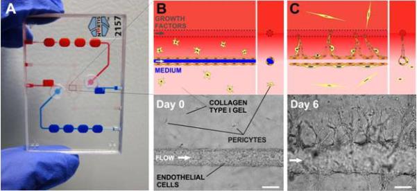
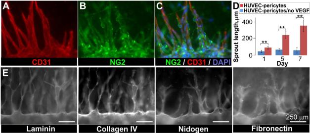
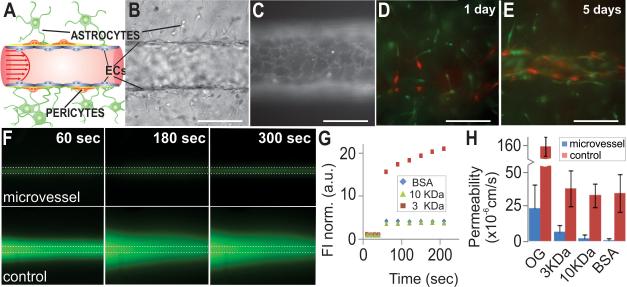
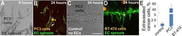
The high attrition rate of drug candidates late in the development process has led to an increasing demand for test assays that predict clinical outcome better than conventional 2D cell culture systems and animal models. Government agencies, the military, and the pharmaceutical industry have started initiatives for the development of novel in-vitro systems that recapitulate functional units of human tissues and organs. There is growing evidence that 3D cell arrangement, co-culture of different cell types, and physico-chemical cues lead to improved predictive power. A key element of all tissue microenvironments is the vasculature. Beyond transporting blood the microvasculature assumes important organ-specific functions. It is also involved in pathologic conditions, such as inflammation, tumor growth, metastasis, and degenerative diseases. To provide a tool for modeling this important feature of human tissue microenvironments, we developed a microfluidic chip for creating tissue-engineered microenvironment systems (TEMS) composed of tubular cell structures. Our chip design encompasses a small chamber that is filled with an extracellular matrix (ECM) surrounding one or more tubular channels. Endothelial cells (ECs) seeded into the channels adhere to the ECM walls and grow into perfusable tubular tissue structures that are fluidically connected to upstream and downstream fluid channels in the chip. Using these chips we created models of angiogenesis, the blood-brain barrier (BBB), and tumor-cell extravasation. Our angiogenesis model recapitulates true angiogenesis, in which sprouting occurs from a "parent" vessel in response to a gradient of growth factors. Our BBB model is composed of a microvessel generated from brain-specific ECs within an ECM populated with astrocytes and pericytes. Our tumor-cell extravasation model can be utilized to visualize and measure tumor-cell migration through vessel walls into the surrounding matrix. The described technology can be used to create TEMS that recapitulate structural, functional, and physico-chemical elements of vascularized human tissue microenvironments in vitro.

Keywords: Microfluidic device; body-on-chip; microenvironment; microphysiological system; microvasculature; organ-on-chip; tissue engineering.


Figures

Figure 1
Figure 1. 3D angiogenesis in TEMS chips
A) Photograph of the microfluidic device, clear circles indicate cell injection septa; B) Two parallel tubes are formed in collagen gel mixed with pericytes (yellow stars): one tube (blue) is populated with HUVECs and a pro-angiogenic cocktail (VEGF/b-FGF/PMA) is flowed through the second tube (red) to create an interstitial gradient in collagen (upper panel, schematic; lower panel, brightfield micrograph); C) A growth factor gradient induces sprouting from the parent vessel towards the gradient (upper panel, schematic; lower panel, micrograph). Scale bars are 125 μm.
Figure 2
Figure 2. Angiogenesis model recapitulates features of the in vivo process
Parent HUVEC vessels were created in collagen mixed with pericytes and exposed to a gradient of growth factors for 11 days. A-C) Co-immunofluorescence indicated that pericytes (NG2, green) were recruited to the parent vessels and associated sprouts (CD31, red; DAPI, blue); D) Sprouting from parent vessels was dependent on the presence of a growth factor gradient: plot of sprout length in the presence and absence of a growth factor gradient, error bars are mean ± 1 standard deviation (N=9 vessels, with VEGF; N=3 vessels, no-VEGF control). Unpaired Student's t-test, 95% confidence interval for VEGF and no-VEGF conditions: t(8)=4.4, p = 0.0022, t(5)=7.8, p = 0.0005, t(6)=7.5, p = 0.0003 for days 1, 5, and 7 respectively. E) Basement membrane proteins were deposited along the parent HUVEC vessel and associated sprouts grown in co-culture with pericytes. Detergent-free immunofluorescence staining. Scale bars are 250 μm.
Figure 3
Figure 3. Paracellular permeability in the BBB model
A) Schematic of the in vivo human BBB*; B) Recapitulation of BBB features: the vessel wall is formed by ECs with pericytes and astrocytes located in the proximity (oblique illumination image); C) CD31 immunostaining shows correct localization of cell junctions and complete cell coverage; D) A microvessel was created from immortalized brain microvascular cells (hCMEC/D3) surrounded by ECM embedded with pericytes (red) and astrocytes (green); E) Within 5 days of culture, the astrocytes and pericytes self-organized to associate with the brain microvessel, no retraction of the endothelium from the walls was observed. Scale bars in (C-E) are 125 μm; F) Brain microvessels and empty channels (control) were perfused with fluorescently tagged BSA and images were taken every 30 sec for 10 min; G) Plot of normalized fluorescent intensity (FI) vs. time in the regions adjacent to the microvessel for molecules of different MW; H) Permeability coefficient for Oregon Green (OG, n=22 vessels, n=3 control tubes), Alexa Fluor dextrans 3 KDa (n=13 vessels, n=3 control tubes), and 10 KDa (n=18 vessels, n=3 control tubes), and BSA-Alexa Fluor 594 (BSA, n=28 vessels, n=3 control tubes). Unpaired Student's t-test, 95% confidence interval, for the permeability of vessels and controls to all four tracers: t(23)=9.3, p< 0.0001, t(14)=7.6, p<0.0001, t(19)=13.7, p<0.0001, t(29)=14.7, p < 0.0001, for OG, 3KDa and 10 KDa dextrans, and BSA, respectively. Error bars are mean ± 1 standard deviation, Scale bars are 250 μm. *modified after: Abbot et al. Nature Reviews Neuroscience (7) 2006, 42-53.
Figure 4
Figure 4. Cancer cell migration through microvessel walls
A) Oblique illumination image of PC3 prostate cancer cells that were introduced into a HUVEC microvessel sprout; note the spherical cells marked with arrows. PC3 and endothelial cells were stained with live cell tracker dyes prior to cell injection; B) Twenty-four hours later the PC3 cells (orange-yellow) had transmigrated through the endothelial cell (green) vessel wall into the surrounding matrix; C) PC3 cells did not transmigrate when injected into empty collagen channels, oblique illumination; D) No transmigration of BT-474 breast cancer cells (orange-yellow) was observed 24 hours after introduction into HUVEC (green) sprouts; E) Plot of the percentage of PC3 and BT-474 cells in HUVEC sprouts that extravasated through the microvessel wall or percentage of PC3 cells in empty channel (control) that migrated into surrounding matrix. Data is plotted as the mean ± 1 standard deviation. (control: n=4 channels (3540 total cells in channels); PC3: n=5 vessels (167 total cells in sprouts); BT-474: n=2 vessels)). Scale bar is 125 μm.

References

    1. Arrowsmith J, Miller P. Trial watch: phase II and phase III attrition rates 2011-2012. Nat Rev Drug Discov. 2013;12:569. - PubMed
    1. Baker BM, Chen CS. Deconstructing the third dimension: how 3D culture microenvironments alter cellular cues. J Cell Sci. 2013;125:3015–24. - PMC - PubMed
    1. Schmeichel KL, Bissell MJ. Modeling tissue-specific signaling and organ function in three dimensions. J Cell Sci. 2003;116:2377–88. - PMC - PubMed
    1. Birgersdotter A, Sandberg R, Ernberg I. Gene expression perturbation in vitro--a growing case for three-dimensional (3D) culture systems. Semin Cancer Biol. 2005;15:405–12. - PubMed
    1. Huh D, Hamilton GA, Ingber DE. From 3D cell culture to organs-on-chips. Trends Cell Biol. 2011;21:745–54. - PMC - PubMed

Publication types

MeSH terms