

Annual bone loss and success rates of dental implants based on radiographic measurements
- PMID: 25030551
- PMCID: PMC4170845
- DOI: 10.1259/dmfr.20140007
Annual bone loss and success rates of dental implants based on radiographic measurements
Abstract
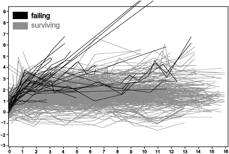
Objectives: Bone loss around dental implants is generally measured by monitoring changes in marginal bone level using radiographs. After the first year of implantation, an implant should have <0.2 mm annual loss of marginal bone level to satisfy the criteria of success. However, the process of measuring marginal bone level on radiographs has a precision of 0.2 mm (or more) owing to variations in exposure geometry, exposure time and observer perception. Therefore, the value of the annual loss may vary considerably, especially when short intervals are considered. This study investigates how the success rate of dental implants depends on the way annual bone loss is calculated.
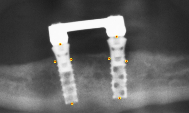
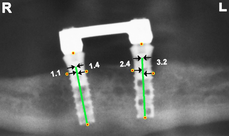
Methods: Panoramic radiographs of 82 implant patients with an average follow-up of 10.4 years were analysed. Marginal bone levels near the implants were indicated by one observer. The annual loss of marginal bone level was determined according to four different calculation methods.
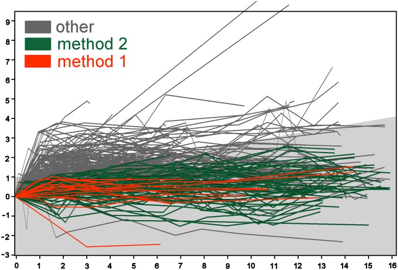
Results: The methods yielded success rates of 9%, 45%, 81% and 89%.
Conclusions: The success rate of dental implants measured on radiographs greatly depends on the details of the calculation method. Without rigorous standardization, annual bone loss and implant success rate are not well defined.
Keywords: criteria; marginal distance; noise; radiographs; success rate.
Figures
References
-
- Esposito M, Hirsch JM, Lekholm U, Thomsen P. Biological factors contributing to failures of osseointegrated oral implants. (I) Success criteria and epidemiology. Eur J Oral Sci 1998; 106: 527–51. - PubMed
-
- Albrektsson T, Zarb G, Worthington P, Eriksson AR. The long-term efficacy of currently used dental implants: a review and proposed criteria of success. Int J Oral Maxillofac Implants 1986; 1: 11–25. - PubMed
-
- Chaytor DV. Clinical criteria for determining implant success: bone. Int J Prosthodont 1993; 6: 145–52. - PubMed
-
- Smith DE, Zarb GA. Criteria for success of osseointegrated endosseous implants. J Prosthet Dent 1989; 62: 567–72. - PubMed
-
- Wyatt CC, Pharoah MJ. Imaging techniques and image interpretation for dental implant treatment. Int J Prosthodont 1998; 11: 442–52. - PubMed
LinkOut - more resources
Full Text Sources
Other Literature Sources
Medical
