. 2014 Jul 17:5:4421.
doi: 10.1038/ncomms5421.

Endocannabinoids modulate cortical development by configuring Slit2/Robo1 signalling

Affiliations

Endocannabinoids modulate cortical development by configuring Slit2/Robo1 signalling

Alán Alpár et al. Nat Commun. .

Abstract

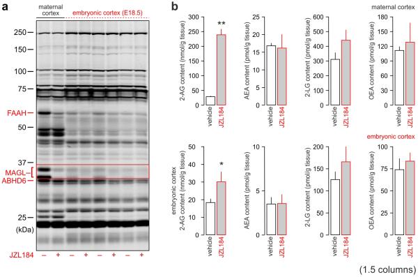
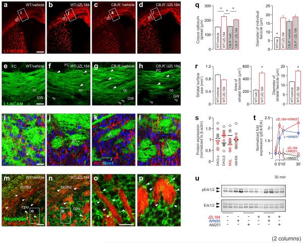
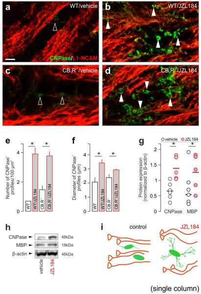
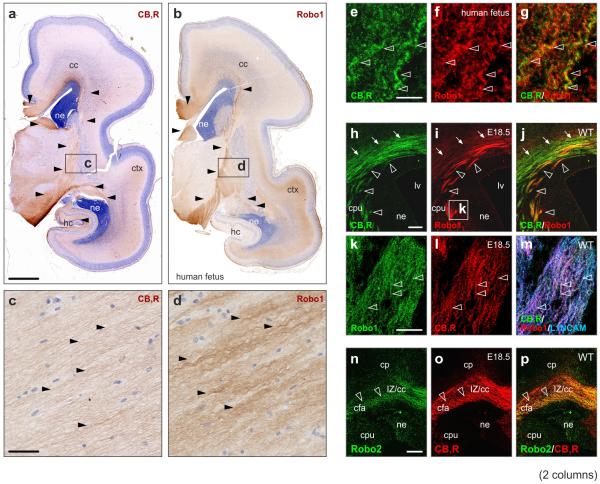
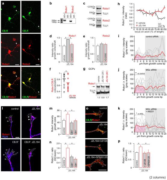
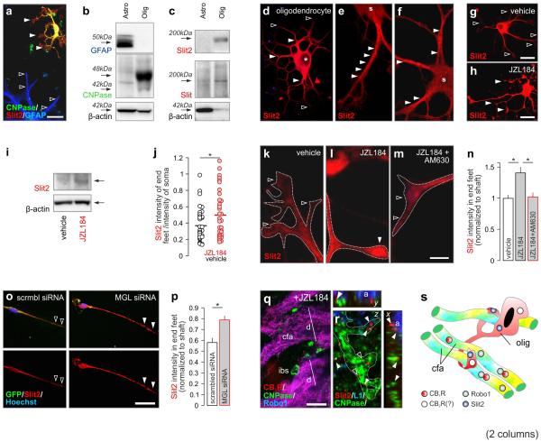
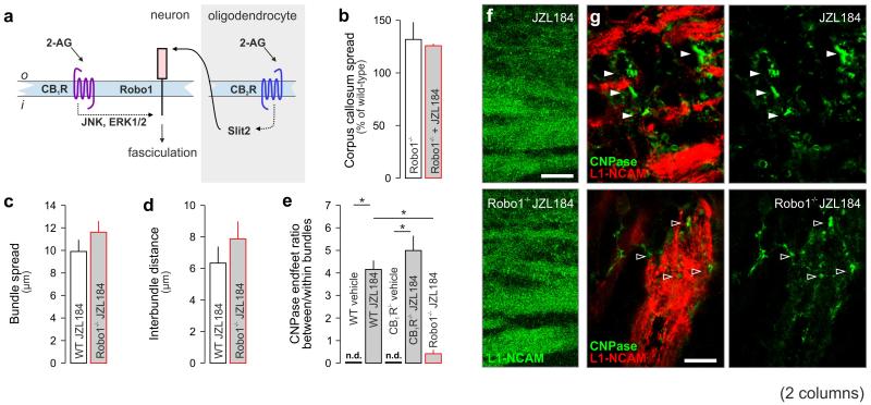
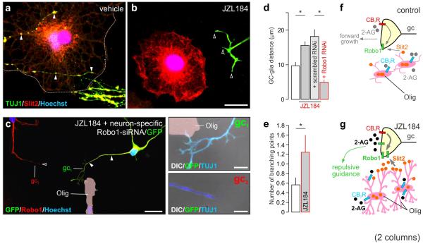
Local environmental cues are indispensable for axonal growth and guidance during brain circuit formation. Here, we combine genetic and pharmacological tools, as well as systems neuroanatomy in human fetuses and mouse models, to study the role of endocannabinoid and Slit/Robo signalling in axonal growth. We show that excess 2-arachidonoylglycerol, an endocannabinoid affecting directional axonal growth, triggers corpus callosum enlargement due to the errant CB1 cannabinoid receptor-containing corticofugal axon spreading. This phenotype mechanistically relies on the premature differentiation and end-feet proliferation of CB2R-expressing oligodendrocytes. We further show the dependence of both axonal Robo1 positioning and oligodendroglial Slit2 production on cell-type-specific cannabinoid receptor activation. Accordingly, Robo1 and/or Slit2 manipulation limits endocannabinoid modulation of axon guidance. We conclude that endocannabinoids can configure focal Slit2/Robo1 signalling to modulate directional axonal growth, which may provide a basis for understanding impaired brain wiring associated with metabolic deficits and prenatal drug exposure.


Figures

Fig. 1
Fig. 1
JZL184 increases 2-AG levels in maternal and embryonic brains. (a) Ex vivo activity-based protein profiling of brain-derived serine hydrolases in vehicle and JZL184 (40 mg/kg)- treated adult and fetal cortices. (b) Select 2-monoacyl glycerol and N-acyl amide levels in maternal and fetal brains correlatively measured after JZL184 treatment. Data were expressed as means ± s.e.m., n = 2/group (mothers), n = 6/group (fetuses); **p < 0.01, *p < 0.05 (Student’s t-test).
Fig. 2
Fig. 2
JZL184 alters callosal axon fasciculation and pathfinding. (a-d) Corticofugal axonal phenotypes and (b-h) their midline crossing in vehicle or JZL184-treated mouse embryos at E18.5. (i-l) A taugfp/gfp reporter mouse was used for the genetic tagging of corticofugal axons.. Axon fasciculation errors present in CB1R−/− mice (k), were further increased by JZL184 (surface area occupied by corticofugal axons (l). (m-p) Neurocan was excluded from enlarged axon fascicles in JZL184-exposed brains. (q) Corpus callosum spread in both wild-type and CB1R−/− mice. (r) Striatal surface size, and the cross-sectional surface area and diameter of axon fascicles in JZL184– or vehicle-treated mice. (s) DAGLα, DAGLβ, MGL and ABHD6 protein levels in JZL184-treated (red circles) vs. control (open circles) cortices on E18.5. (t,u) Erk1/2 phosphorylation upon acute WIN55,212-2 and/or AM251 stimulation in drug naive and JZL184 pre-treated (4 days) neurons. Data were expressed as means ± s.e.m., n = 3/group (q,r). In vitro experiments were performed in duplicates with n ≥ 2 samples/experiments run in parallel. *p < 0.05 (Student’s t-test). Scale bars = 300 μm (a-h, m,n), 10 μm (i-l,o,p).
Fig. 3
Fig. 3
JZL184 induces oligodendrocyte differentiation. CNPase+ oligodendroyctes in the prospective corpus callosum were infrequently detected in wild-type (a) and CB1R−/− (c) embryos (E18.5; open arrowheads). (b,d) JZL184-induced oligodendrocyte differentiation, which progressed in CB1R−/− mice (solid arrowheads pinpoint end-feet). (e,f) Quantitative data on oligodendrocyte morphology. (g,h) CNPase and MBP levels in the cortices of JZL184-treated mouse embryos. (i) Schema of JZL184-induced remodeling of axonal pathfinding upon premature oligodendrocyte differentiation. Data were expressed as means ± s.e.m., n = 5/6 (JZL184/vehicle), *p < 0.05 (Student’s t-test). Scale bar = 4 μm (a).
Fig. 4
Fig. 4
CB1Rs co-localize with Robo1 receptors in human and mouse corticofugal axons. (a,b) The regional distribution of CB1R and Robo1 receptor immunoreactivities in human fetal serial sections (closed arrowheads). (c,d) CB1R and Robo1 receptor immunoreactivities in corticofugal fibers in the internal capsule at high-magnification. (e-g) Co-localization of CB1R and Robo1 receptors in human fetal tissues (internal capsule) by high-resolution laser scanning microscopy (open arrowheads). Robo1 (h-m), as well as Robo2 (n-p), was co-expressed with CB1Rs in mouse callosal axons (arrowheads). Abbreviations: cc, corpus callosum; cfa, corticofugal axons; cp, cortical plate; cpu, caudate putamen; hc, hippocampus; IZ, intermediate zone; ne, neuroepithelium. Scale bars = 3mm (a), 200 μm (h,n), 40 μm, (c), 7 μm (k).
Fig. 5
Fig. 5
CB1R stimulation regulates subcellular Robo1 positioning. (a) Robo1 and Robo2 immunoreactivity in the somata and neurites (including growth cone; arrowheads) of CB1R+ cortical neurons in vitro. (b,c) Cultured cortical neurons expressed both Robo1/Robo2 mRNA and protein. (d) Robo1 and Robo2 (e), fluorescence intensity in growth cones treated with JZL184 or O-2050, a CB1R antagonist. (f) Co-localization coefficient of Robo1 and CB1R in growth cones upon JZL184 exposure. (g) JZL184 induced Robo1 accumulation in growth cones ex vivo in isolated growth cone particles (GCPs). (h) Robo1 immunoreactivity adjacent to the growth cone in JZL184 treated neurons. (i-k) Robo1 immunoreactivity in growth cones upon siRNA-mediated MGL silencing in cultured primary neurons. (l-n) Effect of the genetic ablation of CB1Rs in cortical neurons on Robo1 receptor expression in growth cones after JZL184 or vehicle administration. (o,p) The effect of Erk1/2 (U0126) and JNK1 (SP600125) inhibition on Robo1 distribution. Data were expressed as means ± s.e.m., from triplicate experiments (sample size: 8-25 neurons/group in morphometric analysis), *p < 0.05 (Student’s t-test). Scale bars = 10 μm (a), 5 μm (l,o).
Fig. 6
Fig. 6
JZL184 alters Slit2 expression and localization in oligodendrocytes. (a) Oligodendrocytes (solid arrowheads), but not astrocytes, (open arrowheads) contained Slit2. (b) GFAP and CNPase were used to validate the purity of isolated astrocytes and oligodendrocytes, respectively. (c) Oligodendrocytes, but not astrocytes, contained mature Slit2. An additional antibody raised against a phylogenetically conserved epitope of Drosophila Slit confirmed this finding (Slit). (d-f) Slit2-like immunoreactivity localized to oligodendrocyte somas (s) and processes. Membranous staining pattern is shown in both e and f (arrowheads). Asterisks (*) indicate location of the nucleus. (g,h) Oligodendrocyte differentiation and increased Slit2 immunoreactivity, particularly in end-feet (arrowheads), upon JZL184 exposure. (i) Slit2 protein content in oligodendrocytes after JZL184 treatment. (j) JZL184-induced Slit2 accumulation in oligodendrocyte end-feet as measured by quantitative morphometry. (k-k) Effect of AM630, a CB2R antagonist, on JZL184-induced Slit2 accumulation in oligodendrocyte end-feet (open and closed arrowheads). (o,p) The effect of siRNA-mediated MGL inhibition on Slit2 expression in end-feet (arrowheads). (q) CNPase+ oligodendroglial end-feet in the interbundle space (ibs) and in apposition to enlarged fascicles of Robo1+ corticofugal axons (cfa) in JZL184-treated mouse embryos. (r) Three-dimensional reconstruction of Slit2 in oligodendrocyte end-feet (arrowheads) adjacent to corticofugal axons. (s) Schematic of JZL184-induced remodeling of axonal pathfinding upon premature oligodendrocyte differentiation. Data were expressed as means ± s.e.m., n = 8 - 32 cells/group for each condition in duplicate experiments; *p < 0.05 (Student’s t-test). Scale bars = 10 μm (a,h,o,q), 2 μm (m).
Fig. 7
Fig. 7
JZL184-induced oligodendroglial repositioning in Robo1−/− mice. (a) Schematic overview of 2-AG signaling converging on axonal growth and fasciculation through Slit/Robo signaling. (b) Callosal axon fasciculation in Robo1−/− mice treated with JZL184. Note that untreated Robo1−/− mice presented a fasciculation phenotype. (c,d) Diameter of individual bundles and their interbundle distance upon JZL184 treatment in Robo1−/− embryos. (e-g) Oligodendroglia end-feet infiltrated axonal bundles in JZL184-treated Robo1−/− mice. This was undetected (n.d.) in wild-type or CB1R−/− embryos. Data were expressed as means ± s.e.m., from n = 3-5 embryos/genotype; *p < 0.05 vs. respective wild-type (WT) controls (Student’s t-test). Scale bars =10 μm (f,g).
Fig. 8
Fig. 8
JZL184 induces growth cone repulsion via Slit2/Robo1 signaling. (a) Neurites (arrowheads) contacting oligodendrocytes when co-cultured. (b) Neurites (open arrowheads) excluded from the vicinity of oligodendrocytes upon JZL184 application. Note the intensely Slit2+ peripheral domain of the oligodendrocyte (Olig) shown. (c) A neurite (arrowheads) of a Robo1 siRNA/GFP co-transfected neuron (gc1 in green), but not of a non-transfected neuron (gc2 in red), overcame chemorepulsion. Insets (gc1,gc2) depict high-resolution images of the growth cones shown in (c). (d) Growth cone distances to the most proximal surface of the nearest oligodendrocyte. (e) JZL184 induced branching of neuronal processes in the vicinity of oligodendrocytes in vitro. (f,g) Schema of Slit2/Robo1 signaling downstream from cannabinoid receptors. Data were expressed as means ± s.e.m., triplicate experiments with n = 3 samples run in parallel; n = 6-9 observations/group. *p < 0.05 (Student’s t-test). Scale bars = 8 μm (a1,b), 3 μm (insets).

References

    1. Brose K, et al. Slit proteins bind Robo receptors and have an evolutionarily conserved role in repulsive axon guidance. Cell. 1999;96:795–806. - PubMed
    1. Kolodkin AL, Tessier-Lavigne M. Mechanisms and molecules of neuronal wiring: a primer. Cold Spring Harb. Perspect. Biol. 2011;3 - PMC - PubMed
    1. Hohenester E. Structural insight into Slit-Robo signalling. Biochem. Soc. Trans. 2008;36:251–256. - PubMed
    1. Lopez-Bendito G, et al. Robo1 and Robo2 cooperate to control the guidance of major axonal tracts in the mammalian forebrain. J Neurosci. 2007;27:3395–3407. - PMC - PubMed
    1. Andrews W, et al. Robo1 regulates the development of major axon tracts and interneuron migration in the forebrain. Development. 2006;133:2243–2252. - PubMed

Publication types

MeSH terms