. 2014;63(4):395-401.
doi: 10.1538/expanim.63.395. Epub 2014 Jul 15.

Role of articular disc in condylar regeneration of the mandible

Affiliations

Role of articular disc in condylar regeneration of the mandible

Hidetaka Hayashi et al. Exp Anim. 2014.

Abstract

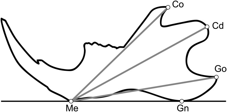
The articular disc in the temporomandibular joint plays an important role in mandibular growth. Functional appliances induce regeneration of the condyle even after condylectomy. The aim of this study was to examine the role of the articular disc in regeneration of the condyle after unilateral condylectomy with use of a functional appliance in growing rats. Fifty growing rats were subjected to unilateral condylectomy and then half of them underwent discectomy. The functional appliance was applied to half of the rats in each group to induce regeneration of the condyle. Four and eight weeks later, morphometric and histologic analyses of the mandible were performed. Regeneration of the condyle was demonstrated in the two condylectomy groups. In the condylectomy+appliance group, the shape and cartilage of the condyle were equivalent to a normal condyle. However, regeneration of the condyle was not observed in the two discectomy groups even with the use of the functional appliance. The articular disc appears to be crucial in the regeneration of a damaged condyle, suggesting that defects or damage to the articular disc may influence mandibular growth and regeneration or repair of the condyle.


Figures

Fig. 1.
Fig. 1.
Landmarks used for cephalometric analyses. Me-Go, Me-Cd, and Me-Co were measured. Me, Point on the most inferior contour of the lower border of the mandible, adjacent to the incisors; Go, Most posterior point of the angular process of the mandible; Cd, Most posterosuperior point of the condylar process; Co, Most posterior point of the coronoid process of the mandible; Gn, Point on the most inferior contour of the angular process of the mandible.
Fig. 2.
Fig. 2.
Micro-CT image of the control and experimental groups at 0, 1, 2, 4, 6 and 8 weeks. In the Cd group, the condyle which was shorter and more irregular than in the control group, was observed at 4, 6 and 8 weeks. In the Cd+Appl group, the condyle which was longer with a more prominent constricted shape of the neck than in the Cd group, was confirmed after 8 weeks by circles. In the Cd+Dc group, only a short process with linear base was seen at 4, 6, or 8 weeks by arrows. In Cd+Dc+Appl group, a tubercle with wide base at the position of condylar process was observed at 4, 6, or 8 weeks by arrow heads.
Fig. 3.
Fig. 3.
H-E stained sections of rat condyle at 0 (control, immediately after condylectomy and immediately after condylectomy with discectomy), 4, and 8 weeks. The area contained within each square is enlarged in the images on the right side (Bar=300 µm).
Fig. 4.
Fig. 4.
Superimposition of mean tracings on the Me-Gn line at Me. (a) Comparison of mandibular measurements in the control, Cd, and Cd+Appl groups at 4 weeks and (b) 8 weeks. (c) Comparison of mandibular measurements in the control, Cd+Dc, and Cd+Dc+Appl groups at 4 weeks and (d) 8 weeks. (e) Size of the mandible in each group at 4 and 8 weeks.
None

References

    1. Bryndahl F., Warfvinge G., Eriksson L., Isberg A.2011. Cartilage changes link retrognathic mandibular growth to TMJ disc displacement in a rabbit model. Int. J. Oral Maxillofac. Surg. 40: 621–627. doi: 10.1016/j.ijom.2011.01.004 - DOI - PubMed
    1. Dörfl J.1980. Migration of tendinous insertions. I. Cause and mechanism. J. Anat. 131: 179–195. - PMC - PubMed
    1. Dörfl J.1980. Migration of tendinous insertions. II. Experimental modifications. J. Anat. 131: 229–237. - PMC - PubMed
    1. Fujita T., Ohtani J., Shigekawa M., Kawata T., Kaku M., Kohno S., Tsutsui K., Tenjo K., Motokawa M., Tohma Y., Tanne K.2004. Effects of sex hormone disturbances on craniofacial growth in newborn mice. J. Dent. Res. 83: 250–254. doi: 10.1177/154405910408300313 - DOI - PubMed
    1. Fujita T., Nakano M., Ohtani J., Kawata T., Kaku M., Motokawa M., Tsuka N., Hayashi H., Tanne K.2010. Expression of Sox 9 and type II and X collagens in regenerated condyle. Eur. J. Orthod. 32: 677–680. doi: 10.1093/ejo/cjp163 - DOI - PubMed

MeSH terms