. 2014 Aug 5;83(6):494-501.
doi: 10.1212/WNL.0000000000000666. Epub 2014 Jul 16.

White matter correlates of cognitive dysfunction after mild traumatic brain injury

Affiliations

White matter correlates of cognitive dysfunction after mild traumatic brain injury

Iain D Croall et al. Neurology. .

Abstract

Objective: To relate neurophysiologic changes after mild/moderate traumatic brain injury to cognitive deficit in a longitudinal diffusion tensor imaging investigation.

Methods: Fifty-three patients were scanned an average of 6 days postinjury (range = 1-14 days). Twenty-three patients were rescanned 1 year later. Thirty-three matched control subjects were recruited. At the time of scanning, participants completed cognitive testing. Tract-Based Spatial Statistics was used to conduct voxel-wise analysis on diffusion changes and to explore regressions between diffusion metrics and cognitive performance.

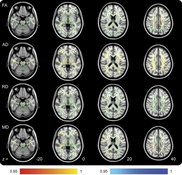
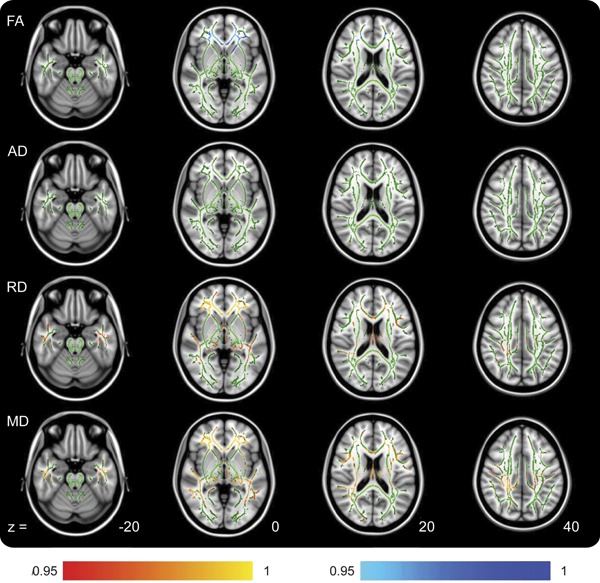
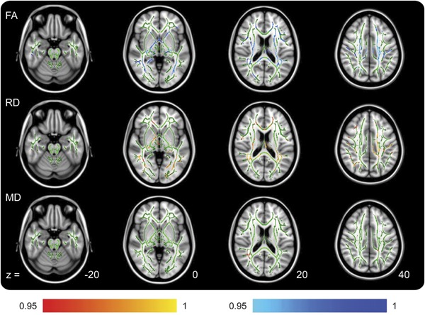
Results: Acutely, increased axial diffusivity drove a fractional anisotropy (FA) increase, while decreased radial diffusivity drove a negative regression between FA and Verbal Letter Fluency across widespread white matter regions, but particularly in the ascending fibers of the corpus callosum. Raised FA is hypothesized to be caused by astrogliosis and compaction of axonal neurofilament, which would also affect cognitive functioning. Chronically, FA was decreased, suggesting myelin sheath disintegration, but still regressed negatively with Verbal Letter Fluency in the anterior forceps.

Conclusions: Acute mild/moderate traumatic brain injury is characterized by increased tissue FA, which represents a clear neurobiological link between cognitive dysfunction and white matter injury after mild/moderate injury.


Figures

Figure 1
Figure 1. TBSS outputs demonstrating locations of metric changes after traumatic brain injury at the acute time point
The analyzed white matter skeleton is shown in green with color overlay of significant differences. All changes are increases (colored red/yellow) in the patient group compared with controls. The z coordinates are based around the anterior commissure/posterior commissure line being z = 0. The RD row shows no differences but is included for comparison with figure 2. Color bar values are 1 − p value. AD = axial diffusivity; FA = fractional anisotropy; MD = mean diffusivity; RD = radial diffusivity; TBSS = Tract-Based Spatial Statistics.
Figure 2
Figure 2. TBSS outputs demonstrating locations of metric changes after traumatic brain injury at the chronic time point
Increases in the patient group are shown in red, and decreases in the patient group are shown in blue. The AD row shows no differences but is included for comparison with figure 1. Color bar values are 1 − p value. AD = axial diffusivity; FA = fractional anisotropy; MD = mean diffusivity; RD = radial diffusivity; TBSS = Tract-Based Spatial Statistics.
Figure 3
Figure 3. TBSS outputs demonstrating locations of regression between acute patient diffusion tensor imaging metrics and Verbal Letter Fluency scores
Positive regressions are shown in red and negative regressions are shown in blue. Axial diffusivity is not shown because no regressions were found for this. Color bar values are 1 − p value. FA = fractional anisotropy; MD = mean diffusivity; RD = radial diffusivity; TBSS = Tract-Based Spatial Statistics.

References

    1. Rugg-Gunn FJ, Symms MR, Barker GJ, Greenwood R, Duncan JS. Diffusion imaging shows abnormalities after blunt head trauma when conventional magnetic resonance imaging is normal. J Neurol Neurosurg Psychiatry 2001;70:530–533 - PMC - PubMed
    1. Goetz P, Blamire A, Rajagopalan B, Cadoux-Hudson T, Young D, Styles P. Increase in apparent diffusion coefficient in normal appearing white matter following human traumatic brain injury correlates with injury severity. J Neurotrauma 2004;21:645–654 - PubMed
    1. Rutgers DR, Fillard P, Paradot G, Tadie M, Lasjaunias P, Ducreux D. Diffusion tensor imaging characteristics of the corpus callosum in mild, moderate, and severe traumatic brain injury. AJNR Am J Neuroradiol 2008;29:1730–1735 - PMC - PubMed
    1. Newcombe V, Chatfield D, Outtrim J, et al. Mapping traumatic axonal injury using diffusion tensor imaging: correlations with functional outcome. PLoS One 2011;6:e19214. - PMC - PubMed
    1. Nakayama N, Okumura A, Shinoda J, et al. Evidence for white matter disruption in traumatic brain injury without macroscopic lesions. J Neurol Neurosurg Psychiatry 2006;77:850–855 - PMC - PubMed

Publication types

LinkOut - more resources