

Geometric Structure of 3D Spinal Curves: Plane Regions and Connecting Zones
- PMID: 25031873
- PMCID: PMC4063218
- DOI: 10.5402/2012/840426
Geometric Structure of 3D Spinal Curves: Plane Regions and Connecting Zones
Abstract
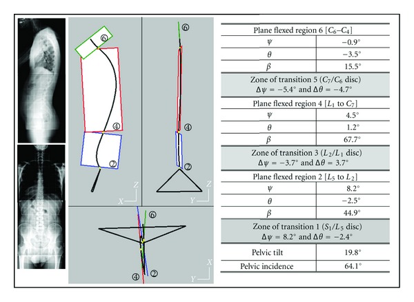
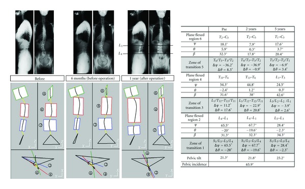
This paper presents a new study of the geometric structure of 3D spinal curves. The spine is considered as an heterogeneous beam, compound of vertebrae and intervertebral discs. The spine is modeled as a deformable wire along which vertebrae are beads rotating about the wire. 3D spinal curves are compound of plane regions connected together by zones of transition. The 3D spinal curve is uniquely flexed along the plane regions. The angular offsets between adjacent regions are concentrated at level of the middle zones of transition, so illustrating the heterogeneity of the spinal geometric structure. The plane regions along the 3D spinal curve must satisfy two criteria: (i) a criterion of minimum distance between the curve and the regional plane and (ii) a criterion controlling that the curve is continuously plane at the level of the region. The geometric structure of each 3D spinal curve is characterized by the sizes and orientations of regional planes, by the parameters representing flexed regions and by the sizes and functions of zones of transition. Spinal curves of asymptomatic subjects show three plane regions corresponding to spinal curvatures: lumbar, thoracic and cervical curvatures. In some scoliotic spines, four plane regions may be detected.
Figures


References
-
- Jackson RP, Peterson MD, McManus AC, Hales C. Compensatory spinopelvic balance over the hip axis and better reliability in measuring lordosis to the pelvic radius on standing lateral radiographs of adult volunteers and patients. Spine. 1998;23(16):1750–1767. - PubMed
-
- Bernhardt M, Bridwell KH. Segmental analysis of the sagittal plane alignment of the normal thoracic and lumbar spines and thoracolumbar junction. Spine. 1989;14(7):717–721. - PubMed
-
- Vedantam R, Lenke LG, Keeney JA, Bridwell KH. Comparison of standing sagittal spinal alignment in asymptomatic adolescents and adults. Spine. 1998;23(2):211–215. - PubMed
-
- Goldberg MS, Poitras B, Mayo NE, Labelle H, Bourassa R, Cloutier R. Observer variation in assessing spinal curvature and skeletal development in adolescent idiopathic scoliosis. Spine. 1988;13(12):1371–1377. - PubMed
LinkOut - more resources
Full Text Sources