Case Reports
. 2014 Mar;4(Suppl 1):S9-S13.
doi: 10.4103/2141-9248.131689.

Trevor Disease (Hemimelic Epiphyseal Displasia): 12-year Follow-up Case Report and Literature Review

Affiliations
Case Reports

Trevor Disease (Hemimelic Epiphyseal Displasia): 12-year Follow-up Case Report and Literature Review

Ds Baumfeld et al. Ann Med Health Sci Res. 2014 Mar.

Abstract

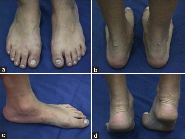
Trevor disease or hemimelic epiphyseal dysplasia is a rare skeletal developmental disorder characterized by asymmetric overgrowth of cartilage in the epiphyses. Histologically, it is an epiphysis osteochondroma. The symptom onset occurs primarily during childhood. Males are 3 times more affected than females. The most common symptom is a painless bony mass around the ankle or knee, followed by swelling, restricted range of motion and deformity. Imaging diagnosis is based on plain radiographs, computed tomography scans and magnetic resonance imaging. Treatment depends on the deformities, symptoms, location and amount of epiphysis involvement. Asymptomatic patients require no treatment. When no deformities are identified, simple mass excision is the treatment choice. If the mass causes epiphyses asymmetry, resection must be combined with osteotomies. The present study reports a case of Trevor disease in a female patient with 12-year follow-up. A general review of Trevor disease was also performed.

Keywords: Ankle; Foot; Foot bones; Foot deformities; Foot diseases.


Conflict of interest statement

Conflict of Interest: None declared.

Figures

Figure 1
Figure 1
Clinical presentation: (a) Dorsal view of the foot. Note blisters at the medial malleolus and I metatarsophalangeal joint; (b) posterior ankle view showing hindfoot valgism with medial protuberance; (c) dorsal foot and ankle views showing hindfoot valgism; (d) posterior ankle view showing hindfoot valgism despite plantar flexion
Figure 2
Figure 2
Left foot X-ray: (a) Anteroposterior view; (b) lateral view; (c) oblique view and (d) axial calcaneus view. Note the gross talar head irregularity with dense areas permeated by radiolucent areas, similar to osteochondroma
Figure 3
Figure 3
Left foot computed tomography and magnetic resonance imaging (MRI) images - gross deformation of the talus head and neck producing articular surface irregularities and hindfoot blocking. Bone features are similar to normal tissue in MRI
Figure 4
Figure 4
(a) Medial aspect of the foot demonstrating skin incision; (b) medial approach showing the bone mass in talus head; (c) resecting bone mass and modeling talo-navicular joint. (d) Bone fragments removed from talo-navicular protuberance
Figure 5
Figure 5
Histopathological blade (H and E): (a) Normal bone tissue covered by normal cartilaginous tissue; (b) set of chondrocytes embedded in chondral matrix within normal bone tissue; (c and d) image enlargement showing cartilaginous islands permeating normal bone
Figure 6
Figure 6
2-year follow-up. (a) Dorsal foot and ankle view; (b) medial aspect of the left foot demonstrating surgical scar; (c) posterior foot and ankle views showing no signs of disease recidivism; (d) heel rise test demonstrating remaining ankle valgism
Figure 7
Figure 7
2 year post-surgery X-ray: (a) Anteroposterior view showing talo-navicular arthrodesis; (b) lateral view showing healed talo-navicular arthrodesis
Figure 8
Figure 8
Clinical presentation 12 years post-surgery. (a) Dorsal foot view; (b) posterior foot and ankle views showing no signs of disease recidivism; (c) medial foot view showing satisfactory scar aesthetics; (d) heel rise test demonstrating the calcaneous remains in valgus
Figure 9
Figure 9
12 years post-surgery X-ray: (a) Anteroposterior foot view with no signs of disease recidivism; (b) lateral foot view demonstrating complete arthrodesis healing

References

    1. Mouchet A, Belot J. La tarsomegalie. J Radiol Electrol. 1926;10:289–93.
    1. Trevor D. Tarso-epiphysial aclasis; a congenital error of epiphysial development. J Bone Joint Surg Br. 1950;32-B:204–13. - PubMed
    1. Fairbank TJ. Dysplasia epiphysialis hemimelica (tarso-ephiphysial aclasis) J Bone Joint Surg Br. 1956;38-B:237–57. - PubMed
    1. Carlson DH, Wilkinson RH. Variability of unilateral epiphyseal dysplasia (dysplasia epiphysealis hemimelica) Radiology. 1979;133:369–73. - PubMed
    1. Kettelkamp DB, Campbell CJ, Bonfiglio M. Dysplasia epiphysealis hemimelica. A report of fifteen cases and a review of the literature. J Bone Joint Surg Am. 1966;48:746–65. - PubMed

Publication types