Review
. 2014 Jul 1;3(7):445-464.
doi: 10.1089/wound.2013.0473.

Epithelialization in Wound Healing: A Comprehensive Review

Affiliations
Review

Epithelialization in Wound Healing: A Comprehensive Review

Irena Pastar et al. Adv Wound Care (New Rochelle). .

Abstract

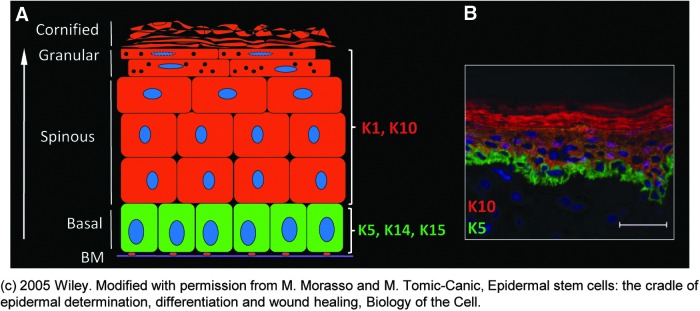
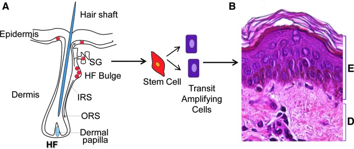
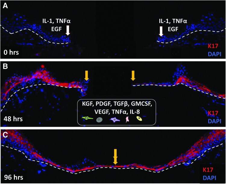
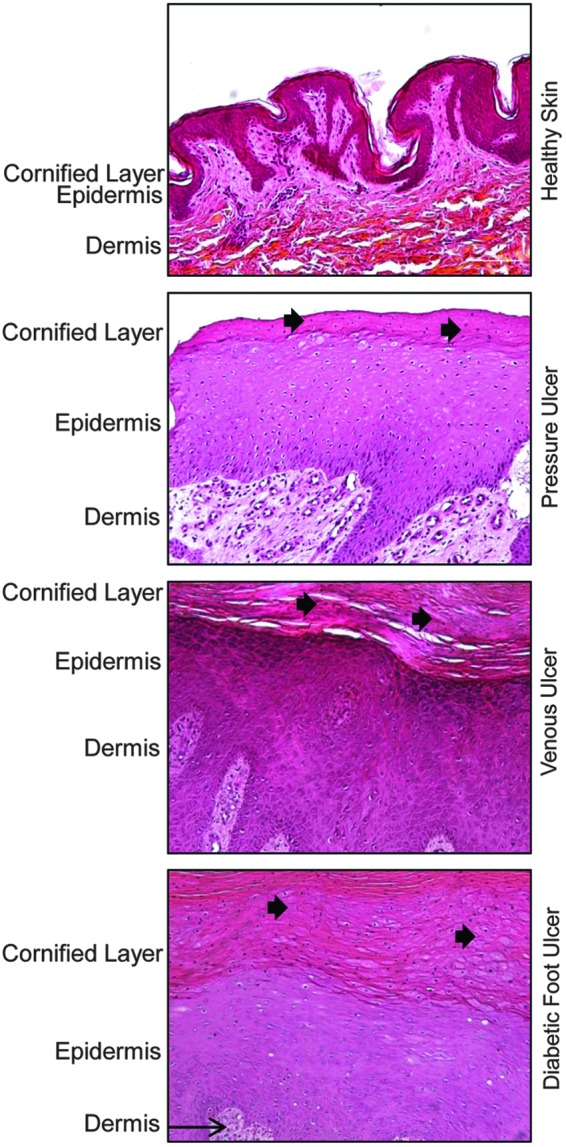
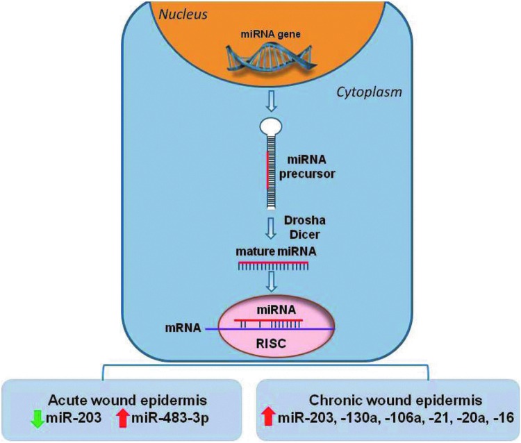
Significance: Keratinocytes, a major cellular component of the epidermis, are responsible for restoring the epidermis after injury through a process termed epithelialization. This review will focus on the pivotal role of keratinocytes in epithelialization, including cellular processes and mechanisms of their regulation during re-epithelialization, and their cross talk with other cell types participating in wound healing. Recent Advances: Discoveries in epidermal stem cells, keratinocyte immune function, and the role of the epidermis as an independent neuroendocrine organ will be reviewed. Novel mechanisms of gene expression regulation important for re-epithelialization, including microRNAs and histone modifications, will also be discussed. Critical Issues: Epithelialization is an essential component of wound healing used as a defining parameter of a successful wound closure. A wound cannot be considered healed in the absence of re-epithelialization. The epithelialization process is impaired in all types of chronic wounds. Future Directions: A comprehensive understanding of the epithelialization process will ultimately lead to the development of novel therapeutic approaches to promote wound closure.


Figures

None
Marjana Tomic-Canic, PhD
<b>Figure 1.</b>
Figure 1.
Structure of the epidermis. (A) Schematic illustration of epidermis. Keratinocytes are the major cell population of epidermis important for maintaining a barrier formation during homeostasis as well as restoring it after the injury. Mitotically active basal layer is adjacent to the basement membrane (BM). Keratinocytes in the basal layer are characterized by keratin (K) 5, K14, and K15. As they differentiate, keratinocytes form suprabasal layers known as the spinous, granular, and cornified layer. Differentiated keratinocytes express K1 and K10. (B) Immunolocalization of K5 (green) and K10 (red) in human epidermis. Nuclei are visualized with 4′,6-diamidino-2-phenylindole–DAPI (blue). © 2005 Wiley. Modified with permission from Morasso and Tomic-Canic. To see this illustration in color, the reader is referred to the web version of this article at www.liebertpub.com/wound
<b>Figure 2.</b>
Figure 2.
Regenerative capacity of the skin relies on local populations of epidermal stem cells. (A) Schematic representation of a hair follicle (HF) with the multipotent stem cells (red). Three distinct epidermal stem cell (ESC) niches are identified so far: bulge of the HF, the base of the sebaceous gland (SG), and the basal layer of the interfollicular epidermis. It has been shown that stem cells migrate from the HF and interfollicular stem cell niche to aid repair and epithelialization upon skin wounding. ESCs generate transit amplifying cells, which will differentiate to form the stratified epidermal layers. (B) Section of human skin stained with hematoxylin and eosin to distinguish epidermis (E) and dermis (D).ORS, outer root sheet; IRS, inner root sheet. To see this illustration in color, the reader is referred to the web version of this article at www.liebertpub.com/wound
<b>Figure 3.</b>
Figure 3.
Epidermal keratinocytes migrate over the wound bed to epithelialize the wound gap. Immunofluorescence staining with keratin 17 (K17, red) antibody demonstrates epithelialization process in human ex vivo wound model. White arrows indicate wound edges after initial wounding, while yellow arrows point at the edges of the migrating epithelial fronts. K17 is not present at the time of wounding (0 h, A). Immediately after injury (A), keratinocytes release proinflammatory cytokines and growth factors, including interleukin 1 (IL-1), tumor necrosis factor α (TNFα), and epidermal growth factor (EGF). In response to these stimuli, keratinocytes become activated and start migrating over the wound bed. Migrating keratinocytes show an upregulation of K17 (48 h, B). Strong K17 staining persisted over 4 days after the wounding when the wound is completely closed (96 h, C). A well-balanced communication with other cell types, fibroblasts, neutrophils, endothelial cells, monocytes, and macrophages (schematically presented at the bottom), B) through various cytokines and growth factors (KGF, PDGF-bb, VEGF, GM-CSF, TGFβ, IL-8), is necessary for successful epithelialization. Nuclei are visualized with DAPI (blue). White dashed lines indicate the dermal–epidermal boundary. KGF, keratinocyte growth factor; PDGF-bb, platelet-derived growth factor bb; VEGF, vascular endothelial growth factor; TGFβ, transforming growth factor β; GM-CSF, granulocyte–macrophage colony-stimulating factor; IL-8, interleukin 8. To see this illustration in color, the reader is referred to the web version of this article at www.liebertpub.com/wound
<b>Figure 4.</b>
Figure 4.
Epidermis of chronic wounds shows specific morphology that differs from epidermis of healthy skin. Healthy human skin is composed of several layers of keratinocytes. These cells lose their nuclei through the process of differentiation and form enucleated cornifed layer. However, epidermis of chronic wounds, such as pressure ulcers, venous ulcers, and diabetic foot ulcers, has distinct morphology. They are characterized by hyperproliferative epidermis as a result of c-myc activation and overexpression. In addition, presence of hyperkeratosis (a thick cornified layer) and parakeratosis [presence of the nuclei in the cornified layer (black arrow)] are additional hallmarks portraying epidermal keratinocytes of chronic wounds. To see this illustration in color, the reader is referred to the web version of this article at www.liebertpub.com/wound
<b>Figure 5.</b>
Figure 5.
Role of miRNAs in epithelialization. Schematic representation of miRNA biogenesis: miRNAs are transcribed in the nucleus as 70-bp precursor products that are processed into the mature ∼22-bp products by the Drosha and Dicer cytoplasmic enzymes. The mature miRNA interacts with the RNA-induced silencing complex (RISC) and binds to complementary sequences in target messenger RNAs (mRNA) leading to downregulation of gene expression. Suppression of miR-203 and induction of miR-483-3p promote keratinocyte migration during normal wound healing process. Induction of miRNA—203, -130a, -106a, 21, -20a, -16 in keratinocytes of nonhealing edges of chronic wounds aids to their pathogenesis by inhibiting epithelialization and wound closure. To see this illustration in color, the reader is referred to the web version of this article at www.liebertpub.com/wound

References

    1. Tomic-Canic M, Komine M, Freedberg IM, and Blumenberg M: Epidermal signal transduction and transcription factor activation in activated keratinocytes. J Dermatol Sci 1998; 17:167. - PubMed
    1. Fuchs E. and Cleveland DW: A structural scaffolding of intermediate filaments in health and disease. Science 1998; 279:514. - PubMed
    1. de Guzman Strong C, Wertz PW, Wang C, Yang F, Meltzer PS, Andl T, et al. : Lipid defect underlies selective skin barrier impairment of an epidermal-specific deletion of Gata-3. J Cell Biol 2006; 175:661. - PMC - PubMed
    1. Eckert RL: Structure, function, and differentiation of the keratinocyte. Physiol Rev 1989; 69:1316. - PubMed
    1. Kalinin AE, Kajava AV, and Steinert PM: Epithelial barrier function: assembly and structural features of the cornified cell envelope. Bioessays 2002; 24:789. - PubMed