

Magnetic resonance imaging following treatment of advanced hepatocellular carcinoma with sorafenib
- PMID: 25032190
- PMCID: PMC4099339
- DOI: 10.3350/cmh.2014.20.2.218
Magnetic resonance imaging following treatment of advanced hepatocellular carcinoma with sorafenib
Abstract
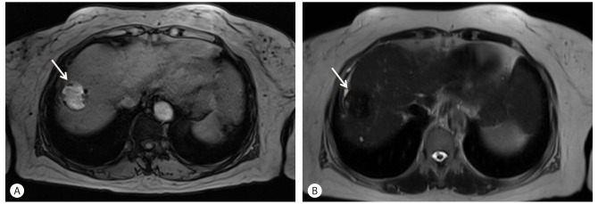
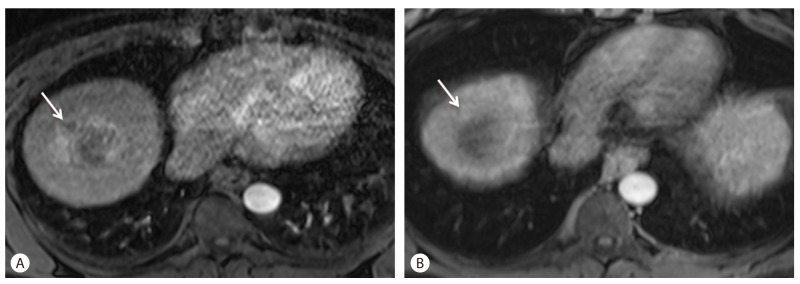
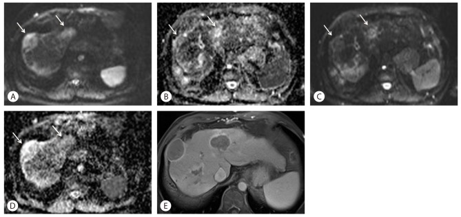
Hepatocellular carcinomas are highly vascular tumors, showing progressive hypervascularity by the process of neoangiogenesis. Tumor angiogenesis is critical for tumor growth as well as metastatic spread therefore, imaging and quantification of tumor neo-angiogenesis is essential for monitoring response to targeted therapies and predicting disease progression. Sorafenib is a molecular targeting agent used for treating hypervascular tumors. This drug is now the standard of care in treatment of patients with advanced hepatocellular carcinoma. Due to its anti-angiogenic and anti-proliferative actions, imaging findings following treatment with Sorafenib are quite distinct when compared to conventional chemotherapeutic agents. Liver MRI is a widely adopted imaging modality for assessing treatment response in hepatocellular carcinoma and imaging features may reflect pathophysiological changes within the tumor. In this mini-review, we will discuss MRI findings after Sorafenib treatment in hepatocellular carcinoma and review the feasibility of MRI as an early biomarker in differentiating responders from non-responders after treatment with molecular targeting agents.
Keywords: Hepatocellular carcinoma; MRI; Sorafenib; mRECIST.
Conflict of interest statement
The authors have no conflicts to disclose.
Figures
References
-
- Kim MJ, Choi JI, Lee JS, Park JW. Computed tomography findings of sorafenib-treated hepatic tumors in patients with advanced hepatocellular carcinoma. J Gastroenterol Hepatol. 2011;26:1201–1206. - PubMed
-
- Lencioni R, Llovet JM. Modified RECIST (mRECIST) assessment for hepatocellular carcinoma. Semin Liver Dis. 2010;30:52–60. - PubMed
-
- Kawaoka T, Aikata H, Murakami E, Nakahara T, Naeshiro N, Tanaka M, et al. Evaluation of the mRECIST and alpha-fetoprotein ratio for stratification of the prognosis of advanced-hepatocellular-carcinoma patients treated with sorafenib. Oncology. 2012;83:192–200. - PubMed
Publication types
MeSH terms
Substances
LinkOut - more resources
Full Text Sources
Other Literature Sources
Medical
