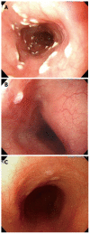
Diagnostic pitfall of sebaceous gland metaplasia of the esophagus
- PMID: 25032211
- PMCID: PMC4097163
- DOI: 10.12998/wjcc.v2.i7.311
Diagnostic pitfall of sebaceous gland metaplasia of the esophagus
Abstract
We investigated the sebaceous gland metaplasia (SGM) of the esophagus and clarified the evidence of misdiagnosis and its diagnosis pitfall. Cases of pathologically proven SGM were enrolled in the clinical analysis and reviewed description of endoscope. In the current study, we demonstrated that SGM is very rare esophageal condition with an incidence around 0.00465% and an occurrence rate of 0.41 per year. There were 57.1% of senior endoscopists identified 8 episodes of SGM. In contrast, 7.7% of junior endoscopists identified SGM in only 2 episodes. Moreover, we investigated the difference in endoscopic biopsy attempt rate between the senior and junior endoscopist (P = 0.0001). The senior endoscopists had more motivation to look for SGM than did junior endoscopists (P = 0.01). We concluded that SGM of the esophagus is rare condition that is easily and not recognized in endoscopy studies omitting pathological review.
Keywords: Endoscopist; Endoscopy; Esophagus; Metaplasia; Sebaceous gland.
Figures


References
-
- Zak FG, Lawson W. Sebaceous glands in the esophagus. First case observed grossly. Arch Dermatol. 1976;112:1153–1154. - PubMed
-
- Nakanishi Y, Ochiai A, Shimoda T, Yamaguchi H, Tachimori Y, Kato H, Watanabe H, Hirohashi S. Heterotopic sebaceous glands in the esophagus: histopathological and immunohistochemical study of a resected esophagus. Pathol Int. 1999;49:364–368. - PubMed
-
- Bambirra EA, de Souza Andrade J, Hooper de Souza LA, Savi A, Ferreira Lima G, Affonso de Oliveira C. Sebaceous glands in the esophagus. Gastrointest Endosc. 1983;29:251–252. - PubMed
-
- Nakada T, Inoue F, Iwasaki M, Nagayama K, Tanaka T. Ectopic sebaceous glands in the esophagus. Am J Gastroenterol. 1995;90:501–503. - PubMed
-
- van Esch E, Brennan S. Sebaceous gland metaplasia in the oesophagus of a cynomolgus monkey (Macaca fascicularis) J Comp Pathol. 2012;147:248–252. - PubMed
Publication types
LinkOut - more resources
Full Text Sources
Other Literature Sources

