

Review
doi: 10.1146/annurev-neuro-071013-013916.
Reassessing models of basal ganglia function and dysfunction
Affiliations
- PMID: 25032493
- PMCID: PMC4416475
- DOI: 10.1146/annurev-neuro-071013-013916
Item in Clipboard
Review
Reassessing models of basal ganglia function and dysfunction
Annu Rev Neurosci.
2014.
Abstract
The basal ganglia are a series of interconnected subcortical nuclei. The function and dysfunction of these nuclei have been studied intensively in motor control, but more recently our knowledge of these functions has broadened to include prominent roles in cognition and affective control. This review summarizes historical models of basal ganglia function, as well as findings supporting or conflicting with these models, while emphasizing recent work in animals and humans directly testing the hypotheses generated by these models.
Keywords: Parkinson's disease; dopamine; striatum.
Figures
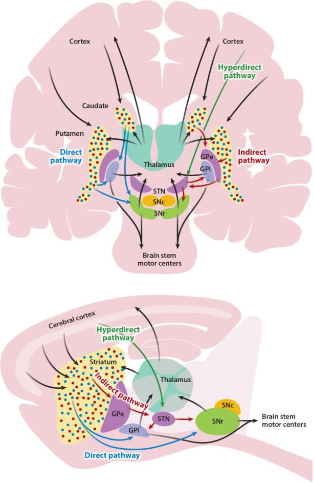
Simplified basal ganglia circuit diagram. Basal ganglia nuclei and their major connections in primates (above), shown in coronal view, and rodents (below), shown in sagittal view. Many additional connections between nuclei are omitted for simplicity; see text for details. In both panels, the direct pathway is shown in blue, the indirect pathway in red, and the hyperdirect pathway in green. Black arrows represent connections shared by multiple pathways. Blue and red dots in the primate caudate/putamen and rodent striatum represent direct pathway–forming and indirect pathway–forming medium spiny neurons, respectively. Abbreviations: GPe, globus pallidus, pars externa; GPi, globus pallidus, pars interna; SNc, substantia nigra, pars compacta; SNr, substantia nigra, pars reticulata; STN, subthalamic nucleus.
References
-
- Albin RL, Young AB, Penney JB. The functional anatomy of basal ganglia disorders. Trends in neurosciences. 1989;12:366–75. - PubMed
-
- Albin RL, Young AB, Penney JB, Handelin B, Balfour R, et al. Abnormalities of striatal projection neurons and N-methyl-D-aspartate receptors in presymptomatic Huntington's disease. The New England journal of medicine. 1990;322:1293–8. - PubMed
-
- Alexander GE, DeLong MR, Strick PL. Parallel organization of functionally segregated circuits linking basal ganglia and cortex. Annual review of neuroscience. 1986;9:357–81. - PubMed
-
- Aylward EH, Sparks BF, Field KM, Yallapragada V, Shpritz BD, et al. Onset and rate of striatal atrophy in preclinical Huntington disease. Neurology. 2004;63:66–72. - PubMed
Publication types
MeSH terms
Grants and funding
LinkOut - more resources
Full Text Sources
Other Literature Sources
