Review
. 2014:37:161-81.
doi: 10.1146/annurev-neuro-071013-014111.

Coupling mechanism and significance of the BOLD signal: a status report

Affiliations
Review

Coupling mechanism and significance of the BOLD signal: a status report

Elizabeth M C Hillman. Annu Rev Neurosci. 2014.

Abstract

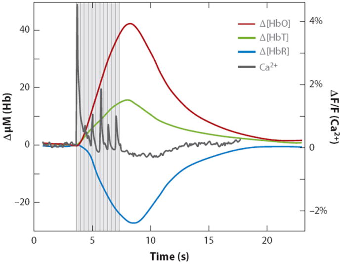
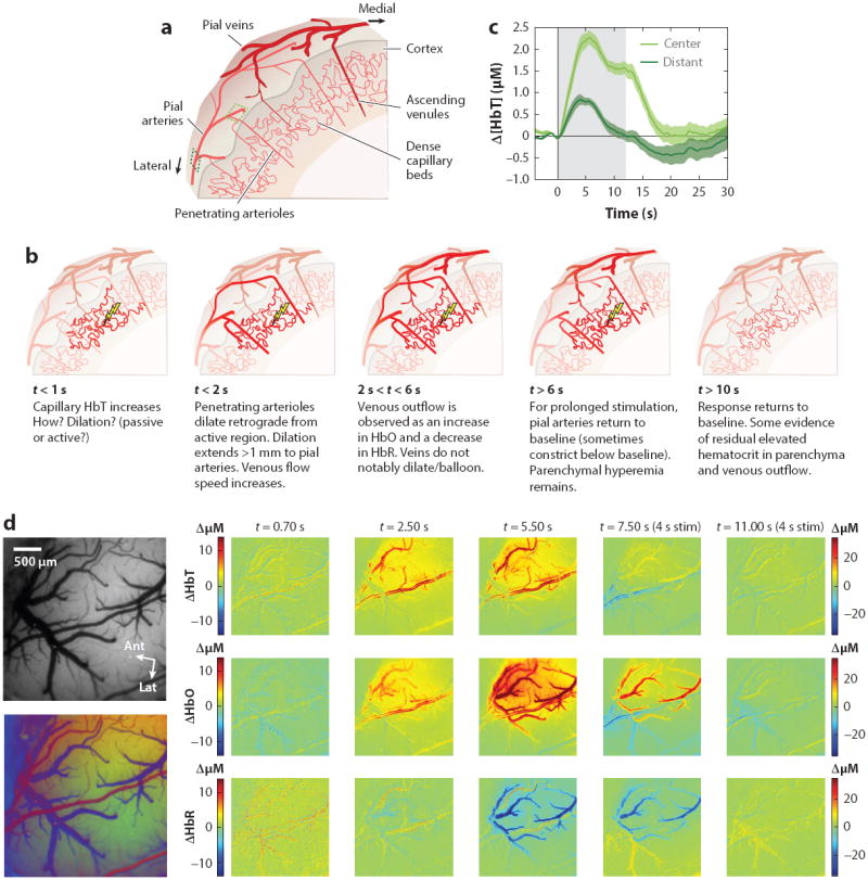
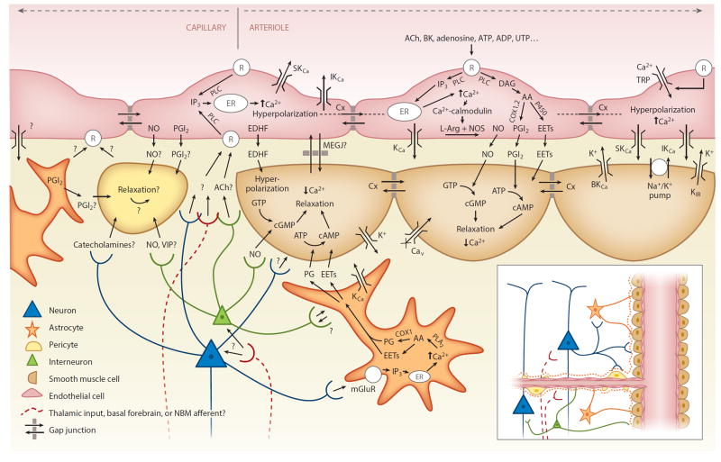
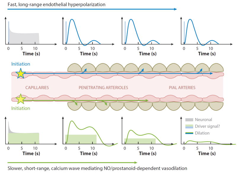
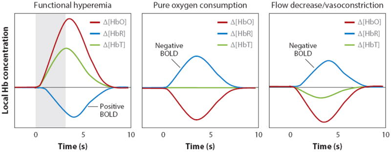
Functional magnetic resonance imaging (fMRI) provides a unique view of the working human mind. The blood-oxygen-level-dependent (BOLD) signal, detected in fMRI, reflects changes in deoxyhemoglobin driven by localized changes in brain blood flow and blood oxygenation, which are coupled to underlying neuronal activity by a process termed neurovascular coupling. Over the past 10 years, a range of cellular mechanisms, including astrocytes, pericytes, and interneurons, have been proposed to play a role in functional neurovascular coupling. However, the field remains conflicted over the relative importance of each process, while key spatiotemporal features of BOLD response remain unexplained. Here, we review current candidate neurovascular coupling mechanisms and propose that previously overlooked involvement of the vascular endothelium may provide a more complete picture of how blood flow is controlled in the brain. We also explore the possibility and consequences of conditions in which neurovascular coupling may be altered, including during postnatal development, pathological states, and aging, noting relevance to both stimulus-evoked and resting-state fMRI studies.

Keywords: astrocytes; fMRI; neurovascular coupling; pericytes; vascular endothelium.


Figures

Figure 1
Figure 1
A typical stimulus-evoked response in the rat somatosensory cortex. Stimulus was 4 s of ~1 mA, 3 Hz forepaw stimulation. Data were acquired using multispectral optical intrinsic signal imaging of the exposed cortex, averaged over the responding region. Dark gray trace shows calcium response to the same stimulation, measured using bulk cortical injection of calcium sensitive fluorophore Oregon green 488 BAPTA-1 AM (Bouchard et al. 2009). Figure reproduced from Hillman (2007). Notably, there is a distinct increase in total hemoglobin (HbT) corresponding to vessel dilation and an increase in the number of red blood cells per unit volume of cortex, consistent with an increase in blood flow. Oxyhemoglobin (HbO) increases while deoxyhemoglobin (HbR) decreases, indicating a net overoxygenation of the region. The fMRI BOLD signal is sensitive to changes in HbR, where stimulus-evoked ‘positive BOLD’ corresponds to the decrease in HbR shown here. The response begins within ~500 ms of stimulus onset and peaks at 3–5 s before slowly returning to baseline. Note that the first large calcium response (corresponding to neuronal activity) precedes marked hemodynamic changes.
Figure 2
Figure 2
Vascular evolution of normal stimulus-evoked functional hyperemia. (a) Schematic cut through of the mammalian cortex. Major cortical blood vessels are located on the pial surface with penetrating arterioles diving perpendicularly into the cortex and branching into dense capillary beds within the cortical layers. Blood drains from these capillaries via perpendicularly oriented ascending venules, which join a network of large draining veins on the pial surface. (b) Schematic sequence of the vascular dynamic response to functional stimulation (see text for citations). (c) Dynamics of the Δ [HbT] response to a 12-s duration, 3 Hz, ~1 mA electrical hindpaw stimulation recorded using optical imaging of the rat somatosensory cortex. ‘Center’ and ‘Distant’ vessels sampled are indicated in panel a. Time courses reveal that the response is more sustained within the central region, while more distant arteries return to baseline earlier, after a peak at 3–5 seconds. Δ [HbT] is independent of oxygenation dynamics and here represents only hyperemia due to arterial/arteriolar dilation and increased concentrations of red blood cells in the capillary beds. (d) A sequence of optical intrinsic signal imaging (OISI) data acquired on the rat somatosensory cortex in response to 4-s, 3 Hz hindpaw stimulation. Left: images showing the field of view under green (530 nm) illumination. Below: a composite based on oxygenation-dependent baseline reflectance highlighting arteries (red), veins (blue), and parenchyma (green). Time sequence: [HbT] changes show an initial increase in parenchymal signal by 0.7 s after stimulus onset (color scale for 0.7 s maps shown at left; all other time points use color scale at right). Increased contrast of pial arteries corresponds to dilation (confirmed by full width half maximum calculation). Increased contrast of the intervening parenchymal space corresponds to an increase in the number of red blood cells per unit volume within the capillary beds (as well as diving and ascending arterioles and venules). [HbR] (deoxyhemoglobin) changes show a distinctly different pattern and can be seen to localize to the shape of the draining veins, with decreases delayed relative to initial changes in [HbT]. Data reproduced from Chen et al. (2011, 2014) and Bouchard et al. (2009).
Figure 3
Figure 3
Candidate neurovascular coupling pathways [modified from Félétou & Vanhoutte (2004) and Attwell et al. (2010)]. Astrocytes can sense glutamate via metabotropic glutamate receptors (mGluR) and increase their intracellular calcium (Ca2+), which can generate arachidonic acid (AA) from phospholipase A2 (PLA2) which is converted by COX1 (or 3) to prostaglandins (PG) and by P450 epoxygenase to epoxyeicosatrienoic acid (EETs). Both PGs and EETs can relax smooth muscle cells (SMCs) through conversion of adenosine triphosphate (ATP) to cyclic adenosine monophosphate (cAMP). Endothelial cells can increase their intracellular calcium through transient receptor potential (TRP) cation channels, and in response to receptor (R) binding, through IP3-mediated release of calcium from intracellular stores [endoplasmic reticulum (ER)]. Endothelial receptor targets include acetylcholine (ACh), bradykinin (BK), adenosine diphosphate (ADP), ATP, uridine triphosphate (UTP), and adenosine. Receptor binding can activate phospholipase C (PLC) (or PLA2), which via diacyl-glycerol (DAG) can also produce EETs and AA derivatives including prostacyclin (PGI2), both of which can drive SMC relaxation via cAMP, while increased intracellular calcium can drive the production of endothelial nitric oxide (NO), which can affect SMC relaxation through conversion of guanosine triphosphate (GTP) to cyclic guanosine monophosphate (cGMP). Intracellular calcium increases also lead to endothelial hyperpolarization through opening of calcium-dependent potassium channels (KCa). Endothelial hyperpolarization could be coupled to adjacent SMCs through myoendothelial gap junctions (MEGJs) or some other endothelium-derived hyperpolarizing factor (EDHF) such as K+ efflux through endothelial SKCa and IKCa channels by activating KIR and/or the Na+/K+ ATPase. SMC hyperpolarization causes relaxation through inactivation of voltage-dependent calcium channels (Cav). Endothelial hyperpolarization can spread rapidly to adjacent endothelial cells, likely via gap junctions. Pericytes possess many SMC-like properties and could relax in response to NO and PGI2 from astrocytes, neurons, or endothelial cells or in response to neuropeptides such as vasointenstinal peptides (VIPs). Pericytes or astrocytes could also be involved in signaling to endothelial cells. Question marks represent many other potential signaling pathways yet to be identified. Additional abbreviation: NMB, nucleus basalis of Meynert.
Figure 4
Figure 4
Proposed model of nonlinear neurovascular coupling incorporating fast and slow propagated vasodilation. Neuronal activity at the capillary level could either directly or indirectly cause an increase in endothelial intracellular calcium. An initial large-amplitude increase in endothelial calcium could initiate endothelial hyperpolarization, which would be rapidly propagated with minimal attenuation to drive relaxation of perivascular SMCs all the way up to the pial arteries (Wölfle et al. 2011). The same initial increase in endothelial calcium could also drive a slower propagating wave of increased calcium within the endothelium (Tallini et al. 2007), bringing NO and prostanoid-dependent vasodilation over a shorter distance. Combined, these two effects would generate spatiotemporal nonlinearities consistent with the properties of functional hyperemia. A lower threshold in endothelial calcium (Marrelli 2001) for slow propagation might explain continued parenchymal hyperemia, but only transient (initial) long-range dilation as shown in Figure 2.
Figure 5
Figure 5
Conditions for positive and negative BOLD. Left: Normal ‘positive BOLD’ in which functional hyperemia increases [HbT] and [HbO]. A decrease in [HbR] occurs because of the wash-in of oxygenated blood. Middle: Possible response when oxygen consumption occurs in the absence of functional hyperemia. This sign of increased metabolic activity would be measured as ‘negative BOLD’. Right: Arteriolar vasoconstriction would decrease [HbT] and [HbO], assuming high arterial oxygen saturation. The consequent decrease in flow would cause deoxygenation, even if oxygen consumption does not change. If there were an associated decrease in the volume of the capillary beds (where oxygen saturation is <98%), the HbR decrease caused by this volume decrease would compete with increasing HbR owing to increasing relative oxygen extraction.

References

    1. Akerboom J, Chen T-W, Wardill TJ, Tian L, Marvin JS, et al. Optimization of a GCaMP calcium indicator for neural activity imaging. J Neurosci. 2012;32:13819–40. - PMC - PubMed
    1. Ances BM, Buerk DG, Greenberg JH, Detre JA. Temporal dynamics of the partial pressure of brain tissue oxygen during functional forepaw stimulation in rats. Neurosci Lett. 2001;306:106–10. - PubMed
    1. Andresen J, Shafi NI, Bryan RM., Jr Endothelial influences on cerebrovascular tone. J Appl Physiol. 2006;100:318–27. - PubMed
    1. Armulik A, Abramsson A, Betsholtz C. Endothelial/pericyte interactions. Circ Res. 2005;97:512–23. - PubMed
    1. Arnerić SP, Honig MA, Milner TA, Greco S, Iadecola C, Reis DJ. Neuronal and endothelial sites of acetylcholine synthesis and release associated with microvessels in rat cerebral cortex: ultrastructural and neurochemical studies. Brain Res. 1988;454:11–30. - PubMed

Publication types