Review
. 2014:37:221-42.
doi: 10.1146/annurev-neuro-062012-170354.

Neural tube defects

Affiliations
Review

Neural tube defects

Nicholas D E Greene et al. Annu Rev Neurosci. 2014.

Abstract

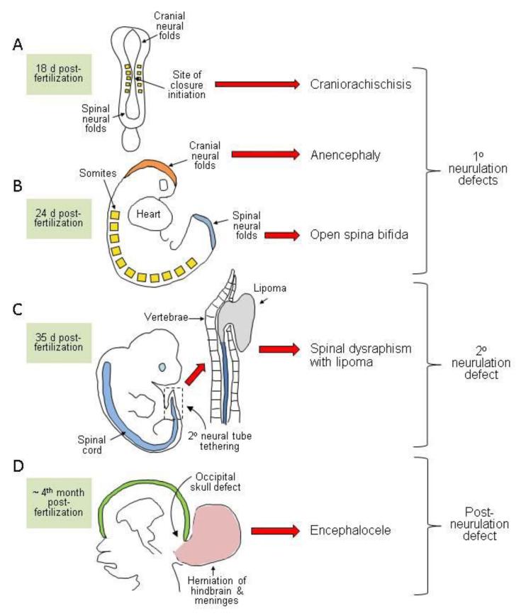
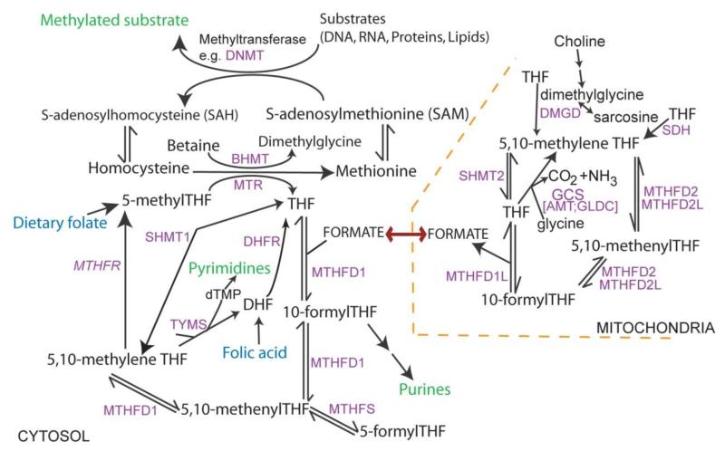
Neural tube defects (NTDs), including spina bifida and anencephaly, are severe birth defects of the central nervous system that originate during embryonic development when the neural tube fails to close completely. Human NTDs are multifactorial, with contributions from both genetic and environmental factors. The genetic basis is not yet well understood, but several nongenetic risk factors have been identified as have possibilities for prevention by maternal folic acid supplementation. Mechanisms underlying neural tube closure and NTDs may be informed by experimental models, which have revealed numerous genes whose abnormal function causes NTDs and have provided details of critical cellular and morphological events whose regulation is essential for closure. Such models also provide an opportunity to investigate potential risk factors and to develop novel preventive therapies.

Keywords: anencephaly; folic acid; genetics; spina bifida.


Figures

Figure 1
Figure 1
Diagrammatic representation of the developmental origin of malformations broadly classified as neural tube defects in humans. (a,b) Disorders of primary neurulation include craniorachischisis (a) in which the neural tube fails to initiate closure, leaving most of the brain and the entire spine open. If closure initiates successfully, then the cranial and/or spinal neural folds may fail to close (b) generating exencephaly/anencephaly and open spina bifida (myelomeningocele), respectively. (c) Disorders of secondary neurulation comprise failure of the neural tube to separate completely from adjacent tissues, resulting in tethering and diminished mobility. The spinal cord is covered by skin and often associated with fatty tissue accumulation (lipoma) through as-yet-unknown mechanisms. (d) Postneurulation defects can arise when the bony structure of the skeleton fails to develop fully. Herniation of the meninges, with or without brain tissue, through a skull defect (shown here as occipital but sometimes parietal or fronto-ethmodial) generates encephalocele, while an analogous defect in the spinal region produces meningocele.
Figure 2
Figure 2. Overview of folate one-carbon metabolism
Folates provide a backbone for the transfer of one-carbon units. Key outputs (in green) include nucleotide biosynthesis and methylation. Among methylation cycle intermediates, homocysteine may also be converted to cystathionine in the transulfuration pathway and S-adenosylmethionine is involved in polyamine biosynthesis. FOCM is compartmentalised: one-carbon units from the mitochondria enter cytoplasmic FOCM as formate while reactions of thymidylate biosynthesis also operate in the nucleus (catalysed by SHMT1, TYMS and DHFR). In loss-of-function mouse models, NTDs arise in mutants for Mthfd1l and genes encoding the glycine cleavage system (GCS). Shmt1 and Mthfr null mice are viable to birth but may develop NTDs under folate-deficient conditions.

References

    1. Abdul-Aziz NM, Turmaine M, Greene ND, Copp AJ. EphrinA-EphA receptor interactions in mouse spinal neurulation: implications for neural fold fusion. Int. J. Dev. Biol. 2009;53:559–68. - PubMed
    1. Adzick NS, Sutton LN, Crombleholme TM, Flake AW. Successful fetal surgery for spina bifida. Lancet. 1998;352:1675–76. - PubMed
    1. Adzick NS, Thom EA, Spong CY, Brock JW, III, Burrows PK, et al. A randomized trial of prenatal versus postnatal repair of myelomeningocele. N. Engl. J. Med. 2011;364:993–1004. - PMC - PubMed
    1. Agopian AJ, Tinker SC, Lupo PJ, Canfield MA, Mitchell LE. Proportion of neural tube defects attributable to known risk factors. Birth Defects Res. A Clin. Mol. Teratol. 2013;97(1):42–46. - PMC - PubMed
    1. Ang S-L, Rossant J. HNF-3β is essential for node and notochord formation in mouse development. Cell. 1994;78:561–74. - PubMed

Publication types

MeSH terms