. 2014 Jul 17;5(7):e1331.
doi: 10.1038/cddis.2014.286.

Ischemic tolerance modulates TRAIL expression and its receptors and generates a neuroprotected phenotype

Affiliations

Ischemic tolerance modulates TRAIL expression and its receptors and generates a neuroprotected phenotype

G Cantarella et al. Cell Death Dis. .

Abstract

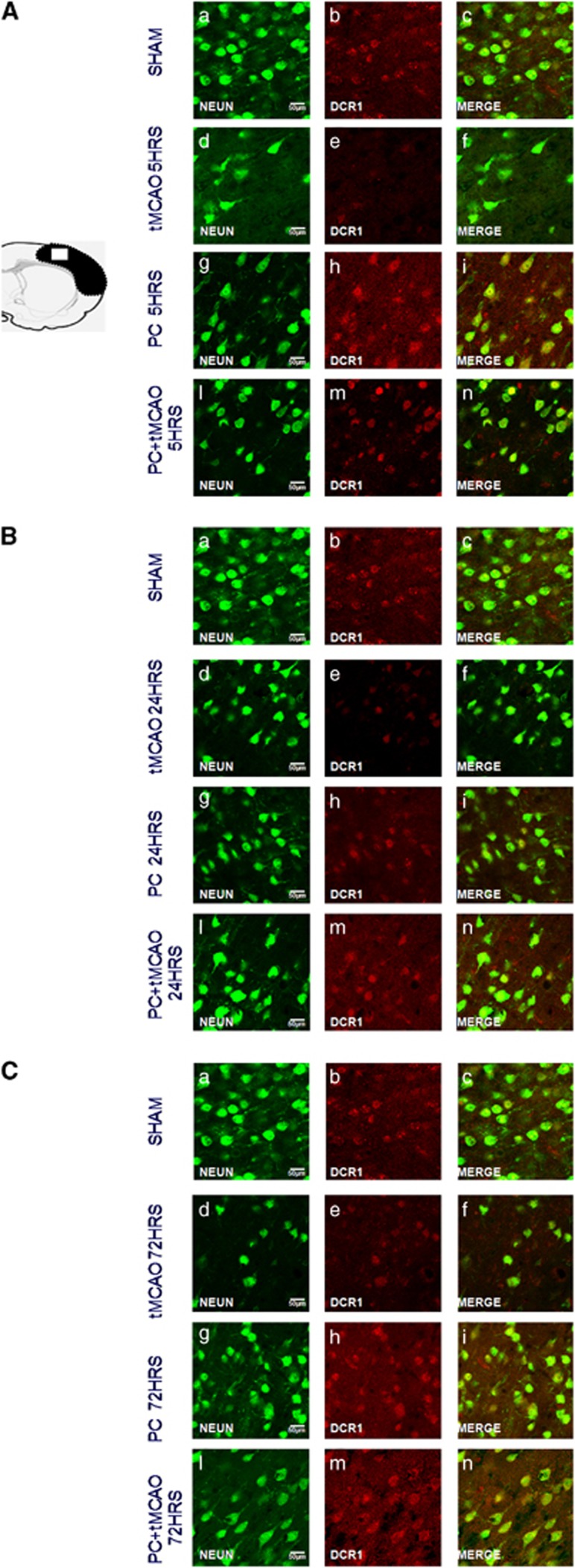
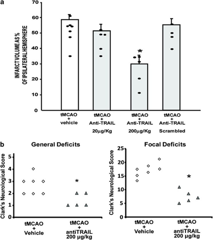
TNF-related apoptosis inducing ligand (TRAIL), a member of the TNF superfamily released by microglia, appears to be involved in the induction of apoptosis following focal brain ischemia. Indeed, brain ischemia is associated with progressive enlargement of damaged areas and prominent inflammation. As ischemic preconditioning reduces inflammatory response to brain ischemia and ameliorates brain damage, the purpose of the present study was to evaluate the role of TRAIL and its receptors in stroke and ischemic preconditioning and to propose, by modulating TRAIL pathway, a new therapeutic strategy in stroke. In order to achieve this aim a rat model of harmful focal ischemia, obtained by subjecting animals to 100 min of transient occlusion of middle cerebral artery followed by 24 h of reperfusion and a rat model of ischemic preconditioning in which the harmful ischemia was preceded by 30 mins of tMCAO, which represents the preconditioning protective stimulus, were used. Results show that the neuroprotection elicited by ischemic preconditioning occurs through both upregulation of TRAIL decoy receptors and downregulation of TRAIL itself and of its death receptors. As a counterproof, immunoneutralization of TRAIL in tMCAO animals resulted in significant restraint of tissue damage and in a marked functional recovery. Our data shed new light on the mechanisms that propagate ongoing neuronal damage after ischemia in the adult mammalian brain and provide new molecular targets for therapeutic intervention. Strategies aimed to repress the death-inducing ligands TRAIL, to antagonize the death receptors, or to activate the decoy receptors open new perspectives for the treatment of stroke.


Figures

Figure 1
Figure 1
Effect of TRAIL administration on neuroprotection elicited by ischemic preconditioning. (a) 6 μg/kg TRAIL i.c.v. administered determines the loss of the neuroprotection elicited by ischemic PC. Infarct volume in rats subjected to tMCAO+Vehicle, tMCAO+0.2 μg/kg TRAIL, tMCAO+6 μg/kg TRAIL, PC+tMCAO+Vehicle, PC+tMCAO+0.2 μg/kg TRAIL, and PC+tMCAO+6 μg/kg TRAIL. Rats were euthanized 24 h after tMCAO. *P<0.05 versus all experimental groups. Each column represents the mean±S.E.M. (n=3–7) of the percentage of the infarct volume compared with the ipsilateral hemisphere. (b) A total of 6 μg/kg TRAIL i.c.v. administered worsens general and focal deficits in rats subjected to PC+tMCAO. *P<0.05 versus PC+vehicle-treated animals
Figure 2
Figure 2
Effect of tMCAO/PC followed by 24 h of reperfusion on either TRAIL and its receptor's protein expression, as well as on TNF-α, FasL, and their respective receptors. (a) Representative blots of DR4, DR5, TRAIL, DcR1, and DcR2 protein expression in sham-operated animals (SHAM) or after 100 min tMCAO (tMCAO), 30 min tMCAO (PC), and 30 min PC, followed, 72 h later, by 100 min tMCAO (PC+tMCAO) in the ipsilateral temporoparietal cortex after 24 h of reperfusion. Tubulin blots: β-tubulin (respective controls). (b) Representative blots of TNFR1, TNF-α, Fas, and FasL protein expression in sham-operated animals (SHAM) and after 100 min tMCAO (tMCAO), 30 min tMCAO (PC), and 30 min PC, followed, 72 h later, by 100 min tMCAO (PC+tMCAO) in the ipsilateral temporoparietal cortex after 24 h of reperfusion. Tubulin blots: β-tubulin (respective controls)
Figure 3
Figure 3
Effect of tMCAO/PC followed by 24 h of reperfusion on either caspases-8 and -3, TRAIL pathway-related kinase JNK, and the cell survival-related kinase Akt. (a) Representative blots of caspase-8 and caspase-3 protein expression in sham-operated animals (SHAM) and after 100 min tMCAO (tMCAO), 30 min tMCAO (PC), and 30 min preconditioning, followed, 72 h later, by 100 min tMCAO (PC+tMCAO) in the ipsilateral temporoparietal cortex after 24 h of reperfusion. Tubulin blots: β-tubulin (controls). (b) Representative blot of phospho-JNK and phospho-AKT protein expression in sham-operated animals (SHAM) or after 100 min tMCAO (tMCAO), 30 min tMCAO (PC), and 30 min PC, followed, 72 h later, by 100 min tMCAO (PC+tMCAO) in the ipsilateral temporoparietal cortex after 24 h of reperfusion. Unphosphorylated JNK and AkT are controls
Figure 4
Figure 4
Effect of 100 min of transient brain ischemia (tMCAO), ischemic PC, and PC followed 72 h later by tMCAO (PC+tMCAO) on TRAIL expression. Confocal microscopic images displaying NeuN (a–l) (green), TRAIL (b–m) (red), and Merge (c–n) (yellow) in the brain peri-ischemic region of rats after 5 h (A), 24 h (B), and 72 h (C) of reperfusion. A representative brain slice cartoon indicating the area of interest is on the left top of the figure. Scale bars in a–i: 50 μm
Figure 5
Figure 5
Effect of 100 min of transient brain ischemia (tMCAO), ischemic PC, and PC followed 72 h later by tMCAO (PC+tMCAO) on DR4 expression. Confocal microscopic images displaying NeuN (a–l) (green), DR4 (b–m) (red), and Merge (c–n) (yellow) in the brain peri-ischemic region of rats after 5 h (A), 24 h (B), and 72 h (C) of reperfusion. A representative brain slice cartoon indicating the area of interest is on the left top of the figure. Scale bars in a–i: 50 μm
Figure 6
Figure 6
Effect of 100 min of transient brain ischemia (tMCAO), ischemic PC, and PC followed 72 h later by tMCAO (PC+tMCAO) on DR5 expression. Confocal microscopic images displaying NeuN (a–l) (green), DR5 (b–m) (red), and Merge (c–n) (yellow) in the brain peri-ischemic region of rats after 5 h (A), 24 h (B), and 72 h (C) of reperfusion. A representative brain slice cartoon indicating the area of interest is on the left top of the figure. Scale bars in a–i: 50 μm
Figure 7
Figure 7
Effect of 100 min of transient brain ischemia (tMCAO), ischemic PC, and PC followed 72 h later by tMCAO (PC+tMCAO) on DcR1 expression. Confocal microscopic images displaying NeuN (A–L) (green), DcR1 (b–m) (red), and Merge (c–n) (yellow) in the brain peri-ischemic region of rats after 5 h (A), 24 h (B), and 72 h (C) of reperfusion. A representative brain slice cartoon indicating the area of interest is on the left top of the figure. Scale bars in a–i: 50 μm
Figure 8
Figure 8
Effect of 100 min of transient brain ischemia (tMCAO), ischemic PC, and PC followed 72 h later by tMCAO (PC+tMCAO) on DcR2 expression. Confocal microscopic images displaying NeuN (a–l) (green), DcR2 (b–m) (red), and Merge (c–n) (yellow) in the brain peri-ischemic region of rats after 5 h (A), 24 h (B), and 72 h (C) of reperfusion. A representative brain slice cartoon indicating the area of interest is on the left top of the figure. Scale bars in a–i: 50 μm
Figure 9
Figure 9
Effect of anti-TRAIL administration on ischemic damage elicited by 100 min of transient middle cerebral occlusion followed by 24 hours of reperfusion. (a) In all, 200 μg/kg antiTRAIL, i.c.v. injected, induces neuroprotection in rats subjected to tMCAO followed by 24 h reperfusion. Infarct volume in rats subjected to tMCAO+Vehicle, tMCAO+20 μg/kg anti-TRAIL, tMCAO+200 μg/kg anti-TRAIL, and tMCAO+200 μg/kg scrambled anti-TRAIL. *P<0.05 versus all experimental groups. Each column represents the mean±S.E.M. (n=5–7) of the percentage of the infarct volume compared with the ipsilateral hemisphere. (b) In all, 200 μg/kg antiTRAIL injected i.c.v. ameliorates general and focal deficits in rats subjected to PC+tMCAO. *P<0.05 versus vehicle-treated animals

References

    1. Lo EH, Dalkara T, Moskowitz MA. Mechanisms, challenges and opportunities in stroke. Nat Rev Neurosci. 4:399–415. - PubMed
    1. Donnan GA, Fisher M, Macleod M, Davis SM. Stroke. Lancet. 2008;371:1612–1623. - PubMed
    1. Muir KW, Tyrrell P, Sattar N, Warburton E. Inflammation and ischaemic stroke. Curr Opin Neurol. 2007;20:334–342. - PubMed
    1. Gidday JM. Cerebral preconditioning and ischaemic tolerance. Nat Rev Neurosci. 2006;7:437–448. - PubMed
    1. Nakajima T, Iwabuchi S, Miyazaki H, Okuma Y, Kuwabara M, Nomura Y, et al. Preconditioning prevents ischemia-induced neuronal death through persistent Akt activation in the penumbra region of the rat brain. J Vet Med Sci. 2004;66:521–527. - PubMed

MeSH terms

Substances