. 2014 Jul 17;5(7):e1332.
doi: 10.1038/cddis.2014.301.

Fluvoxamine alleviates ER stress via induction of Sigma-1 receptor

Affiliations

Fluvoxamine alleviates ER stress via induction of Sigma-1 receptor

T Omi et al. Cell Death Dis. .

Abstract

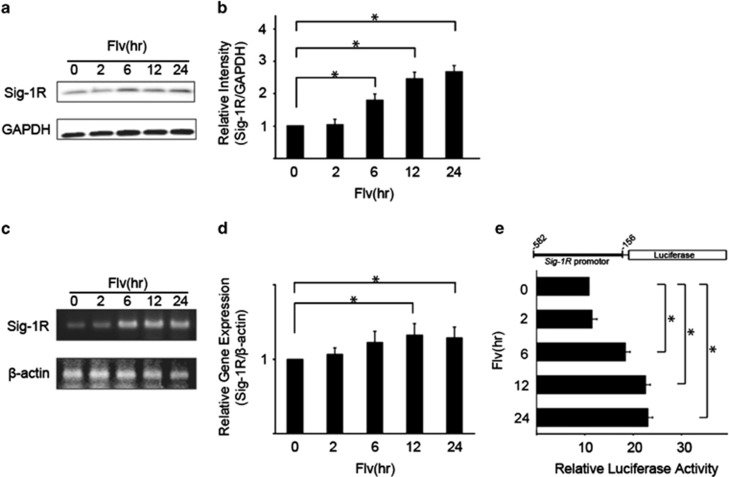
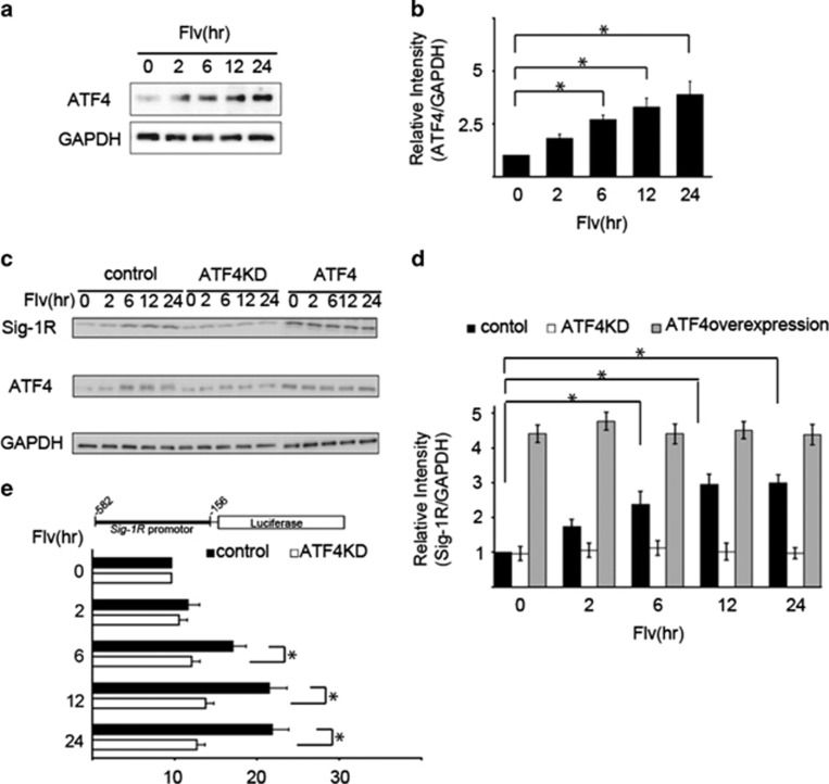
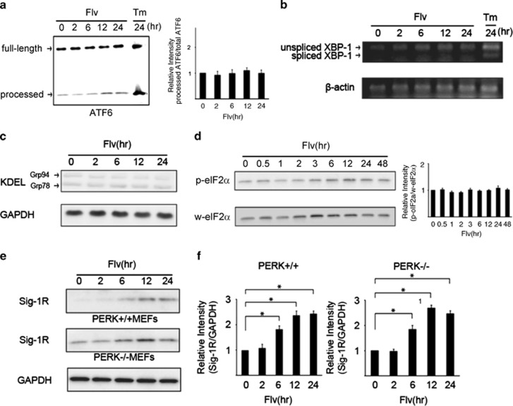
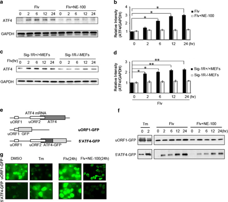
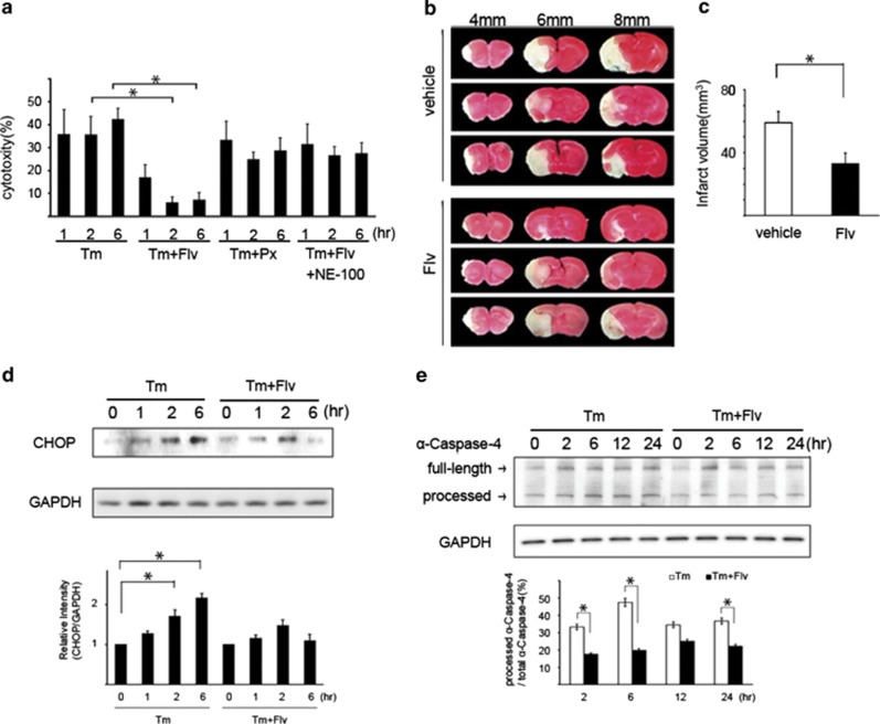
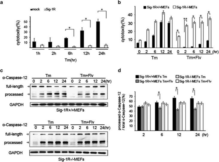
We recently demonstrated that endoplasmic reticulum (ER) stress induces sigma-1 receptor (Sig-1R) expression through the PERK pathway, which is one of the cell's responses to ER stress. In addition, it has been demonstrated that induction of Sig-1R can repress cell death signaling. Fluvoxamine (Flv) is a selective serotonin reuptake inhibitor (SSRI) with a high affinity for Sig-1R. In the present study, we show that treatment of neuroblastoma cells with Flv induces Sig-1R expression by increasing ATF4 translation directly, through its own activation, without involvement of the PERK pathway. The Flv-mediated induction of Sig-1R prevents neuronal cell death resulting from ER stress. Moreover, Flv-induced ER stress resistance reduces the infarct area in mice after focal cerebral ischemia. Thus, Flv, which is used frequently in clinical practice, can alleviate ER stress. This suggests that Flv could be a feasible therapy for cerebral diseases caused by ER stress.


Figures

Figure 1
Figure 1
Fluvoxamine (Flv) induces the expression of sigma-1 receptor (Sig-1R). (a) Flv (10 μg/ml) was added to Neuro2a cells, and Sig-1R protein and GAPDH protein levels were monitored over the time course (h) shown in the figure. (b) The amount of Sig-1R and GAPDH protein in (a) was quantified with densitometry. Relative intensity of Sig-1R/GAPDH increased significantly in response to Flv treatment. The 0 h time point was normalized to 1. Values shown are the mean±S.D. (*P<0.05, Student's t-test; n=3). (c) Sig-1R and β-actin mRNA expression in Neuro2a cells treated with 10 μg/ml Flv was measured by semiquantitative RT-PCR over the time course shown in the figure. The Sig-1R mRNA level increased by ∼3.8-fold after Flv treatment for 24 h. (d) Sig-1R and β-actin mRNA expression in Neuro2a cells treated with 10 μg/ml Flv was determined by quantitative RT-PCR over the time course shown in the figure. The Sig-1R mRNA level increased by 32% and 28% after Flv treatment for 12 h and 24 h, respectively. The 0 h time point was normalized to 1. Values are the mean±S.D. (*P<0.05, Student's t-test; n=3). (e) The reporter assay system shown in the figure was constructed. The Sig-1R promoter region (-582 to -156) was fused to a firefly luciferase plasmid (pGL4.12[luc2CP]) and relative luciferase activities were measured. Luciferase activity increased significantly over the time course shown in response to treatment with 10 μg/ml Flv. Values are the mean±S.D. (*P<0.05, Student's t-test; n=3)
Figure 2
Figure 2
Flv-induced Sig-1R expression is mediated by the transcription factor ATF4. (a) Flv (10 μg/ml) was added to Neuro2a cells, and the expression of ATF4 and GAPDH was measured over the time course shown. (b) The amount of ATF4 and GAPDH protein in (a) was quantified with densitometry. Relative intensity of ATF4/GAPDH increased significantly in response to Flv treatment. The 0 h time point was normalized to 1. Values are the mean±S.D. (*P<0.05, Student's t-test; n=3). (c) Flv (10 μg/ml) was added to HEK293 cells after ATF4 knockdown (ATF4KD), ATF4 overexpression (ATF4), or mock transfection (control). Sig-1R expression was monitored over the time course shown in the figure. In ATF4KD cells, an increase in Sig-1R protein expression was not observed even with Flv treatment. (d) The amount of Sig-1R protein from the samples in panel (c) was quantified with densitometry. Mock-transfected HEK293 cells (control), ATF4KD, and ATF4 HEK293 cells are indicated by black, white, and gray bars, respectively. The 0 h time point was normalized to 1. Values are the mean±S.D. (*P<0.05, Student's t-test; n=3). (e) The reporter assay system shown in the figure was constructed. The Sig-1R promoter region (-582 to -156) was fused to a firefly luciferase plasmid (pGL4.12[luc2CP]) and transfected into HEK293 cells. After 48 h, control cells (control), cells transfected with control shRNA plasmids, and cells transfected with the ATF4-shRNA-pSuper plasmid (ATF4KD) were treated with 10 μg/ml of Flv over the time course shown in the figure. Relative luciferase activities were measured. Sig-1R expression was not significantly induced by Flv treatment in ATF4 knockdown cells. Values are the mean±S.D. (*P<0.05, Student's t-test; n=3)
Figure 3
Figure 3
Flv induces Sig-1R expression without activating the unfolded protein response (UPR) during ER stress. (a) Flv (10 μg/ml) or tunicamycin (Tm; 1 μg/ml) was added to Neuro2a cells, and changes in ATF6α processing were monitored over the time course shown in the figure. The amount of processed ATF6α protein increased with Tm treatment but not with Flv treatment. Processed ATF6α/total ATF6α was measured with densitometry. The results are shown in the bar graph. The 0 h time point was normalized to 1. Values are the mean±S.D. (*P<0.05, Student's t-test; n=3). (b) Flv (10 μg/ml) or Tm (1 μg/ml) was added to Neuro2a cells, and changes in XBP-1 splicing were measured by semiquantitative RT-PCR over the time course shown in the figure. XBP-1 splicing was observed with Tm treatment but not with Flv treatment. (c) Flv (10 μg/ml) was added to Neuro2a cells, and changes in the expression of GRP78/BiP and GRP94, which react with anti-KDEL antibodies, were monitored over the time course shown in the figure. No change in the expression of GRP78/BiP or GRP94 as a result of Flv treatment was observed. (d) Flv (10 μg/ml) was added to Neuro2a cells, and the expression of total eIF2α and phosphorylated eIF2α was measured over the time course shown in the figure. No change in the amount of total eIF2α (w-eIF2α) or phosphorylated eIF2α (p-eIF2α) as a result of Flv treatment was observed. p-eIF2α/w-eIF2α was measured with densitometry. The results are shown in the bar graph. The 0 h time point was normalized to 1. Values are the mean±S.D. (*P<0.05, Student's t-test; n=3). (e) Mouse embryonic fibroblasts (MEFs) from PERK wild-type (PERK+/+) and PERK knockout (PERK−/−) mice were treated with 10 μg/ml of Flv. The expression of Sig-1R and GAPDH was monitored over the time course shown in the figure. Flv treatment induced Sig-1R expression in MEFs from PERK+/+ and PERK−/− animals. (f) Densitometry analysis of (e) showed that relative intensity of Sig-1R/GAPDH increased significantly in both PERK+/+ and PERK−/− MEFs in response to Flv treatment. Left graph, PERK+/+ MEFs; right graph, PERK−/− MEFs. Values are the mean±S.D. (*P<0.05, Student's t-test; n=3)
Figure 4
Figure 4
Flv induces ATF4 via Sig-1R. (a) Neuro2a cells were treated with Flv (10 μg/ml) or Flv (10 μg/ml) and NE-100 (NE-100a Sig-1R antagonist; 1 μg/ml). The expression of ATF4 and GAPDH was monitored over the time course shown in the figure. (b) The amount of ATF4 and GAPDH in (a) was quantified with densitometry. Relative intensity of ATF4/GAPDH in cells treated with Flv+NE-100 was significantly suppressed compared with in cells treated with Flv alone. The 0 h time point was normalized to 1. Values are the mean±S.D. (*P<0.05, Student's t-test; n=3). (c) Flv (10 μg/ml) was added to Sig-1R+/+ MEFs and Sig-1R−/− MEFs. Changes in ATF4 and GAPDH expression were monitored over the time course shown. (d) The amount of ATF4 and GAPDH in (c) was quantified with densitometry. Relative intensity of ATF4/GAPDH showed that ATF4 induction in Sig-1R+/+ MEFs treated with Flv was not observed in Sig-1R−/− MEFs treated with Flv. The 0 h time point was normalized to 1. Values are the mean±S.D. (*P<0.05, **P<0.01, Student's t-test; n=3). (e) To investigate the regulation of ATF4 translation by Flv, we used the reporter assay system shown in the figure. One construct contained ATF4 uORF1 fused with a GFP coding region (uORF1-GFP). A second construct contained a fusion of ATF4 uORF1, uORF2, the ATF4 N terminus, and a GFP coding region (5′ATF4-GFP). (f) HEK293 cells were transfected with the uORF1-GFP reporter construct or the 5′ATF4-GFP reporter construct shown in (e). Thereafter, Tm (1 μg/ml), Flv (10 μg/ml), or Flv (10 μg/ml) and NE-100 (1 μg/ml) were added. Protein expression over the time course shown in the figure was assessed by immunoblot analysis. Changes in GFP expression were monitored. With Tm treatment, GFP expression in uORF1-GFP cells decreased, whereas GFP expression in 5′ATF4-GFP cells increased. On the other hand, with Flv treatment, GFP expression increased in both uORF1-GFP cells and 5′ATF4-GFP cells. The increase in GFP expression observed in uORF1-GFP and 5′ATF4-GFP cells treated with Flv was suppressed when the cells were treated with Flv+NE-100. (g) Cell transfected and treated as described for (f) were observed by fluorescence microscopy. In DMSO-treated cells, uORF1-GFP expression was observed, but 5′ATF4-GFP expression was not detected. In contrast, in Tm-treated cells, uORF1-GFP expression decreased, and 5′ATF4-GFP expression increased, suggesting that ER stress selectively promoted the translation of ATF4. In contrast, increased GFP expression was observed in both uORF1-GFPand 5′ATF4-GFP-transfected cells after Flv treatment (24 h). In both cell lines, the increase in GFP expression was suppressed by treatment with Flv+NE-100 (24 h), consistent with the results of the immunoblot analysis in (f)
Figure 5
Figure 5
Flv suppresses ER stress-mediated apoptosis. (a) Tm, Tm+Flv, Tm+Paroxetine (Px), or Tm+Flv+NE-100 were added to Neuro2a cells. The free LDH activity was measured over the time course shown in the figure, and cytotoxicities were compared. At the times shown in the figure, cells were treated with 10 μg/ml of Flv, 1 μg/ml of Tm, 1 μg/ml of Px, and 1 μg/ml of NE-100. The cytotoxicity induced by Tm+Flv treatment was significantly lower than that induced by treatment with Tm alone. No change in cytotoxicity was observed with Tm+Px treatment. With Tm+Flv+NE-100 treatment, the effect of Flv was negated. Values are the mean±S.D. (*P<0.05, Student's t-test; n=3). (b) Flv administration reduces the area and volume of the cerebral infarct in mice subjected to middle cerebral artery occlusion (MCAO). MCAO was performed in mice administered an intraperitoneal injection of vehicle (20 mg/kg, n=9) or Flv (20 mg/kg, n=9). Cerebral cortex slices were prepared 24 h later. (The forebrain was divided into five coronal 2-mm sections). The slices were stained with TTC, and the areas and volumes of the respective cerebral infarct lesions were compared. The photograph in the figure shows slices 4, 6, and 8 mm from the front of the brain. A significant reduction in the area of the cerebral infarct lesion was observed in Flv-treated mice. Three upper images: representative images of cortex slices from vehicle-treated mice. Three lower images: representative images of cortex slices from Flv-treated mice. (c) MCAO was performed in mice treated with Flv or vehicle. Brain slices from mice in each group were stained with TTC. The area of the cerebral infarct lesion was measured with image analysis software from a photograph of the cut surface taken with a digital camera. Using reported methods, the volume of the cerebral infarct lesion was calculated from the area of the cerebral infarct lesion measured in (b)., The volume of the cerebral infarct lesion in mice treated with Flv was significantly reduced. Values are the mean±S.D. (*P<0.05, Student's t-test; n=3). (d) Tm (1 μg/ml) or Tm (1 μg/ml) and Flv (10 μg/ml) were added to Neuro2a cells, and CHOP expression was monitored over the time course as shown in the figure. A significant reduction in CHOP expression was observed at 2 h and 6 h with Tm+Flv treatment when compared with expression after treatment with Tm alone. CHOP expression was measured with densitometry. The results are shown in the bar graph. The 0 h time point was normalized to 1. Values are the mean±S.D. (*P<0.05, Student's t-test; n=3). (e) Tm (1 μg/ml) or Tm (1 μg/ml) and Flv (10 μg/ml) were added to HEK293 cells, and α-caspase-4 processing was monitored over the time course shown in the figure. Processed α-caspase-4/total α-caspase-4 was measured with densitometry. The results are shown in the bar graph. With the Tm+Flv treatment, α-caspase-4 processing at 2, 6, and 24 h was significantly reduced compared with processing after treatment with Tm alone, indicating that Flv treatment reduced α-caspase-4 activation. Values are the mean±S.D. (*P<0.05, Student's t-test; n=3)
Figure 6
Figure 6
Flv induces neuroprotection by increasing Sig-1R protein expression. (a) Tm (1 μg/ml) was added to mock-transfected HEK293 cells and Sig-1R-overexpressing HEK293 cells. For cytotoxicity comparisons, the free LDH activity was measured over the time course shown in the figure. Cytotoxicity was significantly reduced at 6, 12, and 24 h in Sig-1R expressing cells. Values are the mean±S.D. (*P<0.05, Student's t-test; n=3). (b) Tm (1 μg/ml) or Tm (1 μg/ml) and Flv (10 μg/ml) were added to Sig-1R+/+ and Sig-1R−/− MEFs. The free LDH activity in each treatment condition was measured over the time course shown in the figure, and the cytotoxicities were compared. After Flv treatment, a significant reduction in toxicity was observed in Sig-1R+/+ MEFs but not in Sig-1R−/− MEFs. Values are the mean±S.D. (*P<0.05, Student's t-test; n=3). (c) In the upper figure, Tm (1 μg/ml) or Tm (1 μg/ml) and Flv (10 μg/ml) were added to Sig-1R+/+ MEFs, and caspase-12 processing was monitored over the time course shown. Caspase-12 processing was significantly reduced with Tm+Flv treatment compared with processing after treatment with Tm alone, indicating that Flv suppressed the induction of cell death. In the lower figure, Tm (1 μg/ml) or Tm (1 μg/ml) and Flv (10 μg/ml) were added to Sig-1R−/− MEFs, and caspase-12 processing was monitored over the time course shown. Caspase-12 processing in Sig-1R-deficient cells was not affected by the addition of Flv. (d) Processed caspase-12/total caspase-12 in (c) was measured with densitometry. Black bar: Sig-1R+/+ MEFs treated with Tm; dark gray bar: Sig-1R+/+ MEFs treated with Tm+Flv; light gray bar: Sig-1R−/− MEFs treated with Tm; white bar: Sig-1R−/− MEFs treated with Tm+Flv. Processed caspase-12 was significantly reduced by Flv treatment in Sig-1R+/+ MEF cells but not in Sig-1R−/− MEF cells, demonstrating that the ability of Flv to suppress cell death induction was lost in the absence of Sig-1R. Values are the mean±S.D. (*P<0.05, Student's t-test; n=3)

References

    1. Hayashi T, Su TP. Sigma-1receptor ligands: potential in the treatment of neuropsychiatric disorders. CNS Drugs. 2004;18:269–284. - PubMed
    1. Hayashi T, Su TP. Sigma-1 receptors: evolution of the concept in neuropsychopharmacology. Curr Neuropharmacol. 2005;3:267–280. - PMC - PubMed
    1. Hayashi T, Maurice T, Su TP. Ca2+ signaling via sigma-1 receptors: novel regulatory mechanism affecting intracellular Ca2+concentration. J Pharmacol Exp Ther. 2000;293:788–798. - PubMed
    1. Hayashi T, Su TP. Regulating ankyrin dynamics: Roles of sigma-1 receptors. Proc Natl Acad Sci USA. 2001;98:491–496. - PMC - PubMed
    1. Weissman AD, Casanova MF, Kleinman JE, London ED, De Souza EB. Selective loss of cerebral cortical sigma, but not PCP binding sites in schizophrenia. Biol Psychiatry. 1991;29:41–54. - PubMed