. 2014 Jul 17;9(7):e102860.
doi: 10.1371/journal.pone.0102860. eCollection 2014.

Decay-accelerating factor 1 deficiency exacerbates leptospiral-induced murine chronic nephritis and renal fibrosis

Affiliations

Decay-accelerating factor 1 deficiency exacerbates leptospiral-induced murine chronic nephritis and renal fibrosis

María F Ferrer et al. PLoS One. .

Abstract

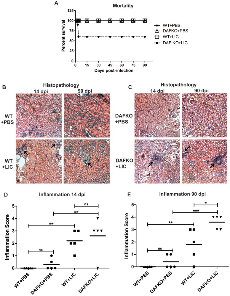
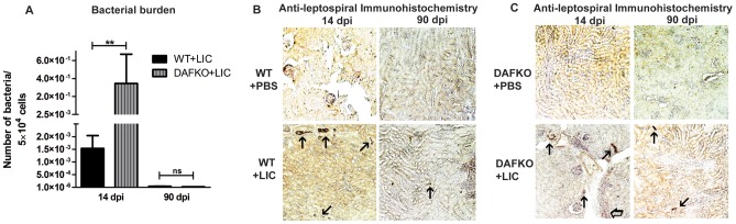
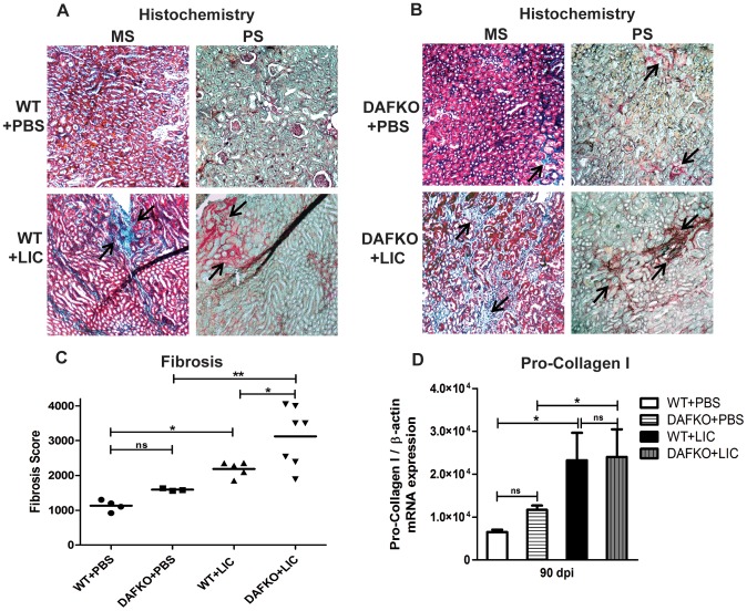
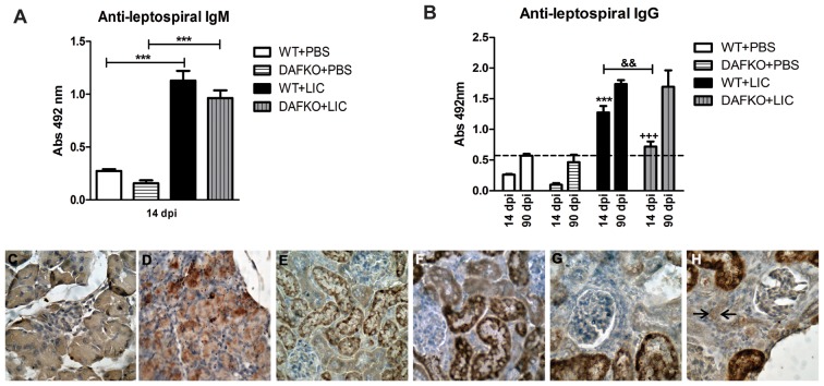
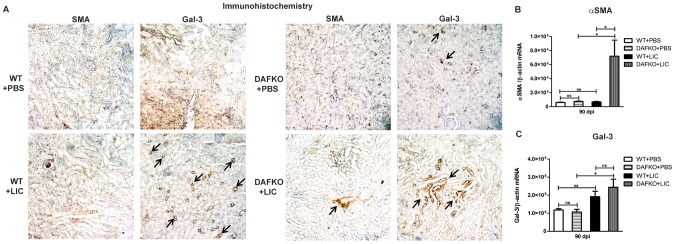
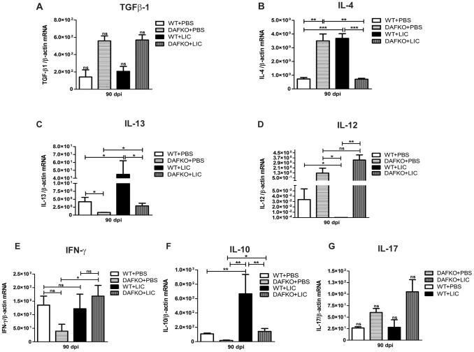
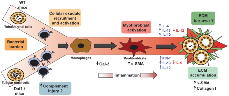
Leptospirosis is a global zoonosis caused by pathogenic Leptospira, which can colonize the proximal renal tubules and persist for long periods in the kidneys of infected hosts. Here, we characterized the infection of C57BL/6J wild-type and Daf1-/- mice, which have an enhanced host response, with a virulent Leptospira interrogans strain at 14 days post-infection, its persistence in the kidney, and its link to kidney fibrosis at 90 days post-infection. We found that Leptospira interrogans can induce acute moderate nephritis in wild-type mice and is able to persist in some animals, inducing fibrosis in the absence of mortality. In contrast, Daf1-/- mice showed acute mortality, with a higher bacterial burden. At the chronic stage, Daf1-/- mice showed greater inflammation and fibrosis than at 14 days post-infection and higher levels at all times than the wild-type counterpart. Compared with uninfected mice, infected wild-type mice showed higher levels of IL-4, IL-10 and IL-13, with similar levels of α-smooth muscle actin, galectin-3, TGF-β1, IL-17, IFN-γ, and lower IL-12 levels at 90 days post-infection. In contrast, fibrosis in Daf1-/- mice was accompanied by high expression of α-smooth muscle actin, galectin-3, IL-10, IL-13, and IFN-γ, similar levels of TGF-β1, IL-12, and IL-17 and lower IL-4 levels. This study demonstrates the link between Leptospira-induced murine chronic nephritis with renal fibrosis and shows a protective role of Daf1.


Conflict of interest statement

Competing Interests: The authors have declared that no competing interests exist.

Figures

Figure 1
Figure 1. Leptospiral-induced nephritis in C57BL/6J wild-type and Daf1−/− mice at 14 and 90 dpi.
A) Survival percentage of WT+PBS (dark rhombus), WT+LIC (white square), DAFKO+PBS (white triangle) and DAFKO+LIC (dark circle) mice. Mice were monitored daily and euthanized by CO2 overdose at 90 dpi (5–7 mice were used). Mortality of C57BL/6J Daf1−/− LIC-infected mice at 3 or 4 dpi was unexpected and presented no previous signs. B–C) Histopathologic sections of uninfected (WT+PBS and DAFKO+PBS) and infected C57BL/6J wild-type (WT+LIC) and Daf1−/− (DAFKO+LIC) kidneys at 14 and 90 dpi assessed by hematoxylin and eosin staining (x200). Lymphomonocytic-rich infiltrates are indicated by arrows. D–E) Inflammation score of uninfected and LIC-infected C57BL/6J wild-type and Daf1−/− animals. Mean inflammation score is represented by a straight line; *p<0.05, **p<0.01, ***p<0.001, ns p>0.05.
Figure 2
Figure 2. Bacterial burden is significantly reduced at later time points.
A) Quantitative measurement of leptospiral DNA (16S) in kidney samples from WT (WT+LIC) or Daf1−/− (DAFKO+LIC) infected animals with 106 bacteria at 14 and 90 dpi. Bars represent standard error mean (SEM) of assays from a group of five to seven mice. Three pieces of each organ were analyzed in triplicate q-PCR and normalized to host cell number; **p<0.01, ns p>0.05. B–C) Immunohistochemistry with antiserum specific for LipL32 (x200) of kidney sections of uninfected (WT+PBS and DAFKO+PBS) and infected WT (WT+LIC) and Daf1−/− (DAFKO+LIC) animals at 14 and 90 dpi. Arrows indicate representative positive foci.
Figure 3
Figure 3. LIC-induced chronic nephritis produces renal fibrosis.
A–B) Renal collagen deposition (indicated by arrows) was analyzed by Masson’s trichrome (MS) and Picro sirius red (PS) staining in uninfected (WT+PBS and DAFKO+PBS) and infected (WT+LIC and DAFKO+LIC) animals at 90 dpi (x200). C) Fibrosis score of WT (WT+PBS and WT+LIC) and Daf1−/− (DAFKO+PBS and DAFKO+LIC) animals. Mean fibrosis score is represented by a straight line, *p<0.05, **p<0.01, ns p>0.05. D) Quantitative measurement of pro-collagen I mRNA in kidney samples from uninfected or infected animals at 90 dpi. Bars represent the standard error of the mean (SEM) of assays from a group of five mice. Three pieces of each organ were analyzed in triplicate for q-PCR and normalized to host β-actin expression; *p<0.05, ns p>0.05.
Figure 4
Figure 4. Anti-leptospiral antibodies followed by enhanced complement activation in Daf1−/− mice suggest a minimal contribution to kidney damage.
A) Total anti-leptospiral IgM from serum samples of uninfected and infected animals at 14 dpi was analyzed by ELISA. Bars represent standard error mean (SEM) of assays from a group of five to seven mice; ***p<0.001. B) Total anti-leptospiral IgG from serum samples of uninfected and infected animals at 14 and 90 dpi was analyzed by ELISA. Bars represent the SEM of assays from a group of five to seven mice; ***p<0.001 with respect to WT+PBS at 14 dpi; +++ p<0.001 with respect to DAFKO+PBS at 14 dpi; && p<0.01 between both WT and Daf1−/− LIC-infected groups. Values under the baseline (dashed line) are considered negligible. Immunohistochemistry with antiserum specific for MAC (membrane attack complex) at 90 dpi (×200). C) Uninfected pancreas as the negative control, D) coxsackievirus B3-infected pancreas as the positive control, WT+LIC without (E) or with anti-MAC (F), DAFKO+LIC without (G) or with anti-MAC (H). Only in panel H there is minimal, positive labeling in interstitial cells indicated by arrows. Samples were treated according to Abcam’s suggested protocol including unmasking. Unmasking usually strongly increases the staining of kidney acinar cells which are rich in endogenous peroxidase, but this staining is intracytoplasmic.
Figure 5
Figure 5. Chronic fibrosis in Daf1−/− +LIC mice induces myofibroblast activation and enhanced galectin-3 expression.
A) Immunohistochemistry of kidney sections from uninfected (WT+PBS and DAFKO+PBS) and LIC-infected (WT+LIC and DAFKO+LIC) mice with antiserum specific for α-smooth muscle actin (α-SMA) or galectin-3 (Gal-3) at 90 dpi (x200). Arrows indicate foci of antigen expression. Quantitative measurement of α-SMA (B) and Gal-3 (C) mRNA expression in kidney samples from uninfected (WT+PBS and DAFKO+PBS) and infected (WT+LIC and DAFKO+LIC) animals at 90 dpi. Bars represent the SEM of assays from a group of five to seven mice. Three pieces of each organ were analyzed in triplicate for q-PCR and normalized to host β-actin expression; *p<0.05, ns p>0.05.
Figure 6
Figure 6. Cytokine levels in renal interstitial fibrosis triggered by LIC infection.
Quantitative measurement of TGF-β1 (A), IL-4 (B), IL-13 (C), IL-12 (D), IFN-γ (E), IL-10 (F) and IL-17 (G) mRNA expression in kidney samples from uninfected (WT+PBS and DAFKO+PBS) or infected (WT+LIC and DAFKO+LIC) animals at 90 dpi. Bars represent the SEM of assays from a group of five to seven mice. Three pieces of each organ were analyzed in triplicate for q-PCR and normalized to host β-actin expression; *p<0.05, **p<0.01, ***p<0.001, ns p>0.05.
Figure 7
Figure 7. Model of leptospiral-induced murine chronic nephritis and renal fibrosis.
Upon LIC colonization of proximal renal tubules of either C57BL/6J wild-type (WT) or Daf1−/− mice, cellular exudate recruitment and activation occurs and precedes the arrival of macrophages, which after stimulation produce mediators such as galectin-3 (Gal-3) that activate quiescent fibroblasts and convert them into an α-SMA+ myofibroblast population. Myofibroblasts produce several molecules and orchestrate the production of ECM components and their extracellular assembly. In WT mice, down-regulation of IL-12 as well as up-regulation of IL-4, IL-10 and profibrotic cytokines such as IL-13 contributes to ECM accumulation. Of note, LIC-infected Daf1−/− mice have a higher bacterial burden at the acute stage of infection, but barely detectable bacterial burden, increased IL-13, IL-10 and IFN-γ expression and decreased IL-4 expression during the chronic stages; these correlate with chronic inflammation, ECM deposition and renal fibrosis. Also, an alternative possibility is that the increased adaptive immune response against infection leads to the deposition of anti-leptospiral antibody followed by complement activation, which then contributes to kidney damage.

References

    1. Sugimoto H, LeBleu VS, Bosukonda D, Keck P, Taduri G, et al. (2012) Activin-like kinase 3 is important for kidney regeneration and reversal of fibrosis. Nat Med 18: 396–404. - PMC - PubMed
    1. Liu Y (2011) Cellular and molecular mechanisms of renal fibrosis. Nat Rev Nephrol 7: 684–696. - PMC - PubMed
    1. Coresh J, Selvin E, Stevens LA, Manzi J, Kusek JW, et al. (2007) Prevalence of chronic kidney disease in the United States. JAMA 298: 2038–2047. - PubMed
    1. Jha V, Garcia-Garcia G, Iseki K, Li Z, Naicker S, et al. (2013) Chronic kidney disease: global dimension and perspectives. Lancet 382: 260–272. - PubMed
    1. Faine S, Adler B, Bolin C, Perolat P (1999) Leptospira and Leptospirosis. Melbourne, Australia MediSci.

Publication types

MeSH terms