. 2014 Jul 17;9(7):e102264.
doi: 10.1371/journal.pone.0102264. eCollection 2014.

Endothelial arginine resynthesis contributes to the maintenance of vasomotor function in male diabetic mice

Affiliations

Endothelial arginine resynthesis contributes to the maintenance of vasomotor function in male diabetic mice

Ramesh Chennupati et al. PLoS One. .

Abstract

Aim: Argininosuccinate synthetase (ASS) is essential for recycling L-citrulline, the by-product of NO synthase (NOS), to the NOS substrate L-arginine. Here, we assessed whether disturbed arginine resynthesis modulates endothelium-dependent vasodilatation in normal and diabetic male mice.

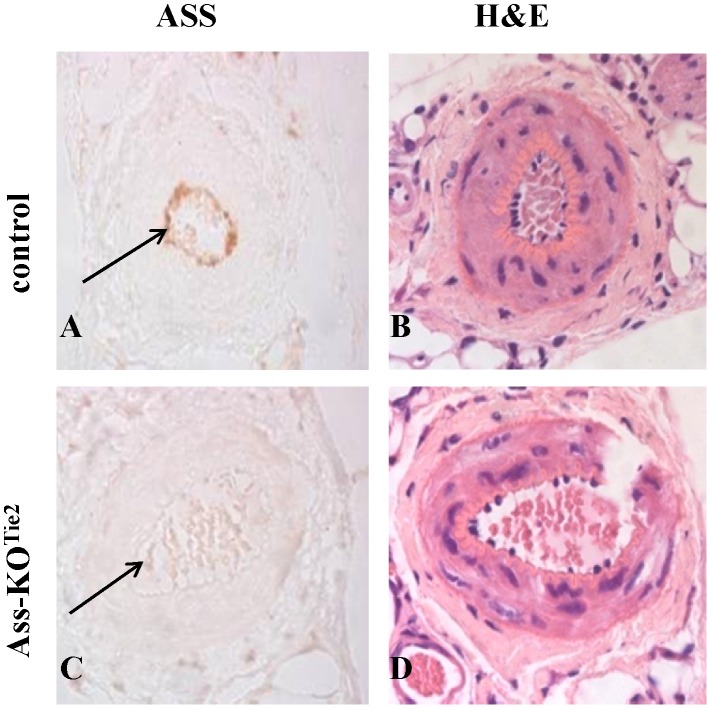
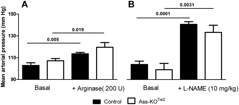
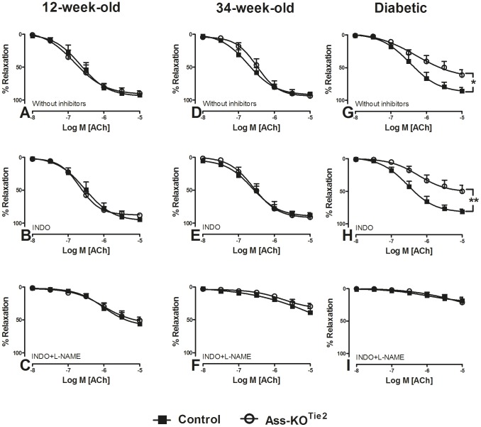
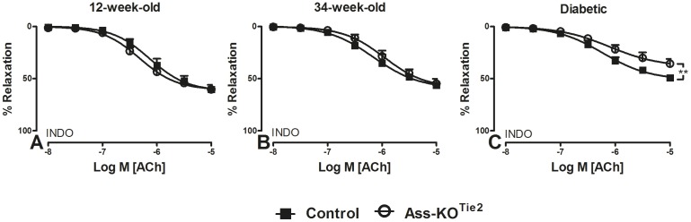
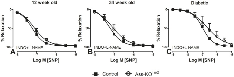
Methods and results: Endothelium-selective Ass-deficient mice (Assfl/fl/Tie2Cretg/- = Ass-KOTie2) were generated by crossing Assfl/fl mice ( = control) with Tie2Cre mice. Gene ablation in endothelial cells was confirmed by immunohistochemistry. Blood pressure (MAP) was recorded in 34-week-old male mice. Vasomotor responses were studied in isolated saphenous arteries of 12- and 34-week-old Ass-KOTie2 and control animals. At the age of 10 weeks, diabetes was induced in control and Ass-KOTie2 mice by streptozotocin injections. Vasomotor responses of diabetic animals were studied 10 weeks later. MAP was similar in control and Ass-KOTie2 mice. Depletion of circulating L-arginine by arginase 1 infusion or inhibition of NOS activity with L-NAME resulted in an increased MAP (10 and 30 mmHg, respectively) in control and Ass-KOTie2 mice. Optimal arterial diameter, contractile responses to phenylephrine, and relaxing responses to acetylcholine and sodium nitroprusside were similar in healthy control and Ass-KOTie2 mice. However, in diabetic Ass-KOTie2 mice, relaxation responses to acetylcholine and endothelium-derived NO (EDNO) were significantly reduced when compared to diabetic control mice.

Conclusions: Absence of endothelial citrulline recycling to arginine did not affect blood pressure and systemic arterial vasomotor responses in healthy mice. EDNO-mediated vasodilatation was significantly more impaired in diabetic Ass-KOTie2 than in control mice demonstrating that endothelial arginine recycling becomes a limiting endothelial function in diabetes.


Conflict of interest statement

Competing Interests: The authors have declared that no competing interests exist.

Figures

Figure 1
Figure 1. Expression of ASS protein in saphenous arteries of male control (panel A) and Ass-KOTie2 (panel C) mice.
The arrow indicates endothelial expression in control animals (panel A) and absence of endothelial expression in Ass-KOTie2 mice (panel C). Panels B and D represent the corresponding H&E staining of a serial section to demonstrate the presence of intact endothelium.
Figure 2
Figure 2. The effect of endothelium-specific Ass deletion on hemodynamics of 34-week-old conscious male mice.
Black bar: control mice; white bar: Ass-KOTie2 mice. Blood pressure was measured in the same mice 2 (panel A) and 3 days (panel B) after catheterization via a femoral artery catheter connected to a pressure transducer. Panel A: mean arterial pressure (MAP) in the basal condition (left) and after a bolus infusion of 200 U bovine arginase 1 via a jugular vein catheter (right). Panel B: mean arterial pressure in the basal condition (left) and after intravenous L-NAME (10 mg/kg) infusion (right). Values are means ± SEM (control animals: arginase 1: n = 7, L-NAME: n = 5; Ass-KOTie2 mice: arginase 1: n = 5, L-NAME: n = 4; due to loss of catheter patency, numbers were lower on the 3rd day). Note that the Y-axis starts at 90 mm Hg.
Figure 3
Figure 3. The effect of endothelium-specific Ass deletion on relaxation responses of saphenous arteries of healthy and diabetic male mice.
Relaxation of PHE (10 µM)-pre-contracted saphenous arteries of 12- (panels A–C) and 34-week-old (panels D–F) healthy and 22-week-old diabetic (panels G–I) male mice to ACh (0.01–10 µM) was determined by wire myography. Black squares: control mice; white circles: Ass-KOTie2 mice. Panels (A, D, G): relaxation responses in the absence of pharmacological inhibitors. Panels (B, E, H): relaxation responses in the presence of INDO (10 µM). Panels (C, F, I): relaxation responses in the presence of both INDO (10 µM) and L-NAME (100 µM). Values are shown as means ± SEM (n = 5–7; for the number of animals per individual experiment see Table 1). *P<0.05 vs. the control, **P<0.01 vs. the control (unpaired t-test).
Figure 4
Figure 4. The effect of endothelium-specific Ass deletion on relaxation responses of saphenous arteries of healthy and diabetic male mice.
Relaxation of K+ (40 mM)-pre-contracted saphenous arteries of 12- (panel A) and 34-week-old (panel B) healthy and 22-week-old diabetic (panel C) male mice to ACh (0.01–10 µM) was determined by wire myography. Black squares: control mice; white circles: Ass-KOTie2. All arteries were treated with INDO (10 µM). Values are shown as means ± SEM (n = 4–8; for the number of animals per individual experiment, see Table 1). **P<0.01 vs. control (unpaired t-test).
Figure 5
Figure 5. The effect of endothelium-specific Ass deletion on relaxation responses of saphenous arteries to sodium nitroprusside.
Relaxation of PHE pre-contracted (10 µM) saphenous arteries of 12- (panel A) and 34-week-old healthy (panel B) and 22-week-old diabetic (C) male mice to SNP (0.01–10 µM) was determined by wire myography. Black squares: control mice; white circles: Ass-KOTie2. All experiments were performed in the presence of L-NAME (100 µM) and INDO (10 µM). Values are means ± SEM (n = 5–7; for the number of animals per individual experiment, see Table 1).

References

    1. Tsihlis ND, Oustwani CS, Vavra AK, Jiang Q, Keefer LK, et al. (2011) Nitric oxide inhibits vascular smooth muscle cell proliferation and neointimal hyperplasia by increasing the ubiquitination and degradation of UbcH10. Cell Biochem Biophys 60: 89–97. - PMC - PubMed
    1. Radomski MW, Palmer RM, Moncada S (1987) Endogenous nitric oxide inhibits human platelet adhesion to vascular endothelium. Lancet 2: 1057–1058. - PubMed
    1. Wang GR, Zhu Y, Halushka PV, Lincoln TM, Mendelsohn ME (1998) Mechanism of platelet inhibition by nitric oxide: in vivo phosphorylation of thromboxane receptor by cyclic GMP-dependent protein kinase. Proc Natl Acad Sci U S A 95: 4888–4893. - PMC - PubMed
    1. Hogg N, Kalyanaraman B, Joseph J, Struck A, Parthasarathy S (1993) Inhibition of low-density lipoprotein oxidation by nitric oxide. Potential role in atherogenesis. FEBS Lett 334: 170–174. - PubMed
    1. Taddei S, Virdis A, Mattei P, Ghiadoni L, Sudano I, et al. (1996) Defective L-arginine-nitric oxide pathway in offspring of essential hypertensive patients. Circulation 94: 1298–1303. - PubMed

Publication types

MeSH terms