Comparative Study
. 2014 Aug 5;53(30):4990-9.
doi: 10.1021/bi500632a. Epub 2014 Jul 24.

Differential activation of intracellular versus plasmalemmal CB2 cannabinoid receptors

Affiliations
Comparative Study

Differential activation of intracellular versus plasmalemmal CB2 cannabinoid receptors

G Cristina Brailoiu et al. Biochemistry. .

Abstract

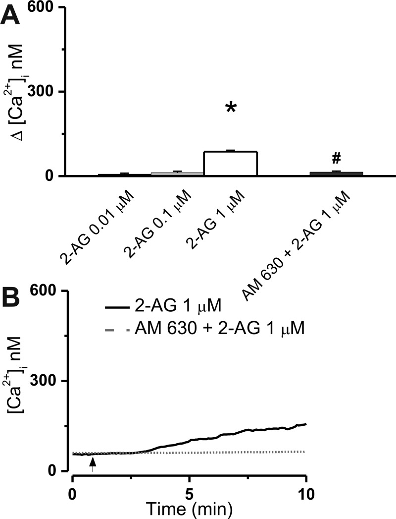
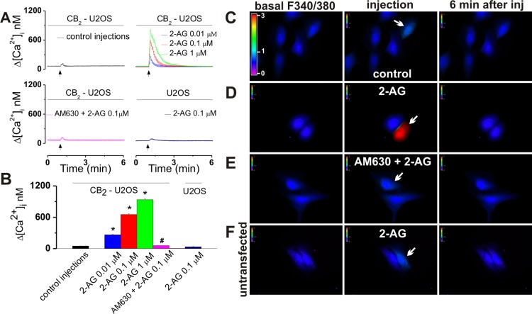
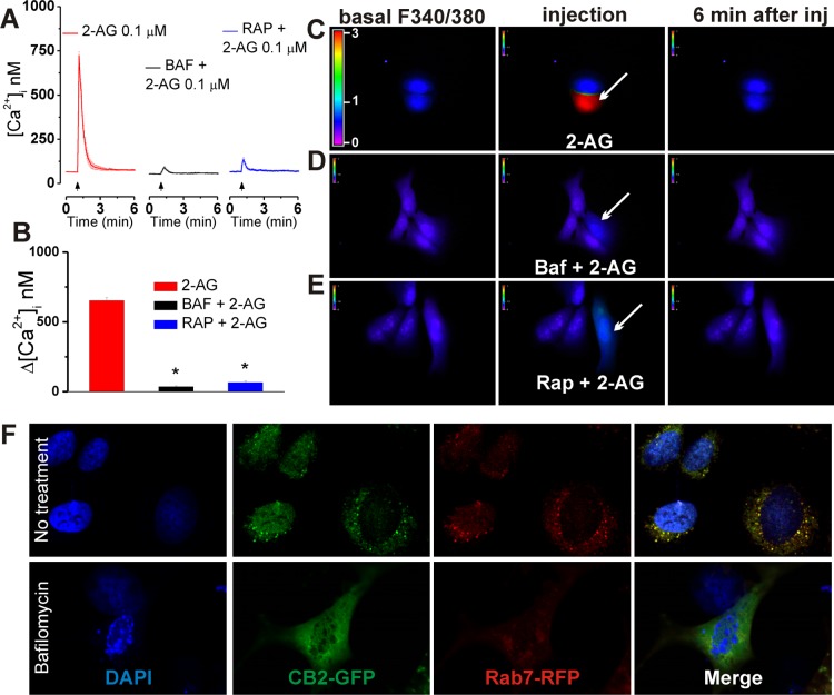
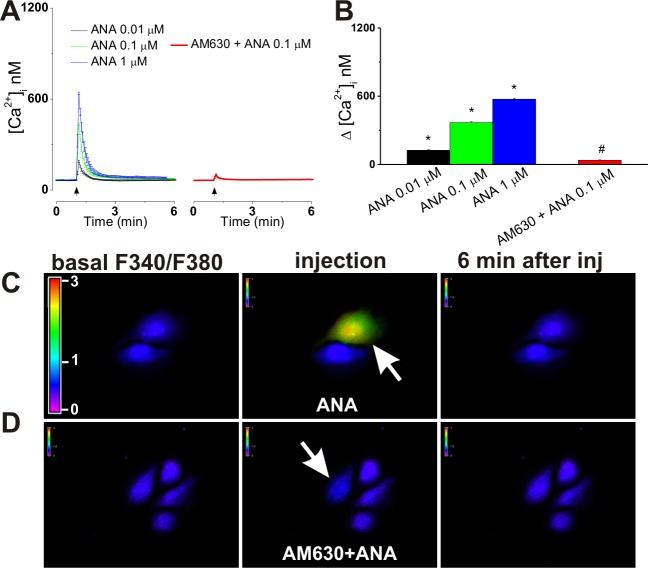
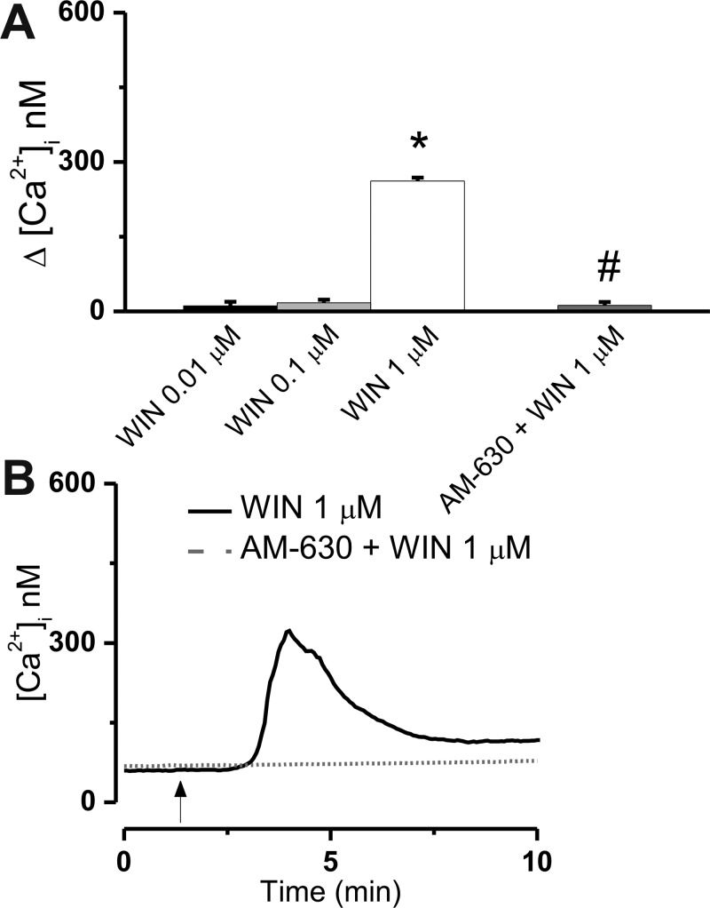
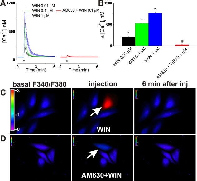
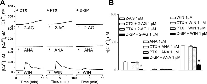
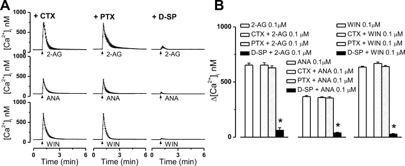
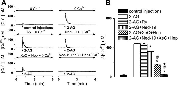
The therapeutic and psychoactive properties of cannabinoids have long been recognized. The type 2 receptor for cannabinoids (CB2) has emerged as an important therapeutic target in several pathologies, as it mediates beneficial effects of cannabinoids while having little if any psychotropic activity. Difficulties associated with the development of CB2-based therapeutic agents have been related to its intricate pharmacology, including the species specificity and functional selectivity of the CB2-initiated responses. We postulated that a plasmalemmal or subcellular location of the receptor may contribute to the differential signaling pathways initiated by its activation. To differentiate between these two, we used extracellular and intracellular administration of CB2 ligands and concurrent calcium imaging in CB2-expressing U2OS cells. We found that extracellular administration of anandamide was ineffective, whereas 2-arachidonoyl glycerol (2-AG) and WIN55,212-2 triggered delayed, CB2-dependent Ca(2+) responses that were Gq protein-mediated. When microinjected, all agonists elicited fast, transient, and dose-dependent elevations in intracellular Ca(2+) concentration upon activation of Gq-coupled CB2 receptors. The CB2 dependency was confirmed by the sensitivity to AM630, a selective CB2 antagonist, and by the unresponsiveness of untransfected U2OS cells to 2-AG, anandamide, or WIN55,212-2. Moreover, we provide functional and morphological evidence that CB2 receptors are localized at the endolysosomes, while their activation releases Ca(2+) from inositol 1,4,5-trisphosphate-sensitive- and acidic-like Ca(2+) stores. Our results support the functionality of intracellular CB2 receptors and their ability to couple to Gq and elicit Ca(2+) signaling. These findings add further complexity to CB2 receptor pharmacology and argue for careful consideration of receptor localization in the development of CB2-based therapeutic agents.


Figures

Figure 1
Figure 1
Extracellular administration of 2-arachidonoyl glycerol (2-AG) to CB2-U2OS cells elevates [Ca2+]i. (A) Comparison of the increases in [Ca2+]i produced by extracellular administration of 2-AG (0.01–1 μM) and 1 μM 2-AG in the presence of CB2 receptor antagonist AM630 (1 μM); P < 0.05 compared with basal levels (∗) or with 1 μM 2-AG (#). (B) Representative recordings of increases in [Ca2+]i in response to 1 μM 2-AG in absence or presence of 1 μM CB2 antagonist AM630.
Figure 2
Figure 2
Intracellular microinjection of 2-AG into CB2-U2OS cells produces CB2-dependent Ca2+ elevation. (A) Averaged increases in [Ca2+]i produced by intracellular administration of control buffer (top left) or 2-AG (0.01, 0.1, and 1 μM, final concentrations inside the cell, top right), co-injection of 0.1 μM 2-AG with CB2 antagonist AM630 (1 μM, bottom left) in CB2-U2OS cells, or microinjection of 0.1 μM 2-AG into control U2OS cells (bottom right). (B) Comparison of the Ca2+ responses elicited by the treatments of CB2-U2OS or control untransfected U2OS cells mentioned above; P < 0.05 when compared with the control (∗) or with 2-AG alone (#). (C–F) Characteristic fluorescence images of Fura-2 AM-loaded CB2-U2OS cells before (left), during (middle), and 6 min after (right) intracellular administration of control buffer (C), 0.1 μM 2-AG alone (D), or 0.1 μM 2-AG in the presence of 1 μM AM630 (E) or of U2OS cells treated with 0.1 μM 2-AG (F). Arrows denote the injected cells; the fluorescence scale (0–3) is illustrated in each panel and magnified in the left panel of part C.
Figure 3
Figure 3
Intracellular CB2 receptors are located at endolysosomes in CB2-expressing U2OS cells. (A) Averaged Ca2+ responses of CB2-U2OS cells to intracellular administration of 0.1 μM 2-AG in the absence (left) or presence of lysosomal disruptor bafilomycin A1 (1 μM, incubation for 1 h, middle) or microautophagy inhibitor rapamycin (30 μM, incubation for 1 h, right). (B) Comparison of the increases in [Ca2+]i elicited by 2-AG under the conditions described above; P < 0.05 when compared with 2-AG microinjection (∗). (C–E) Representative fluorescence images of Fura-2 AM-loaded CB2-U2OS cells before (left), during (middle), and 6 min after (right) intracellular administration of 0.1 μM 2-AG in the absence (C) and presence of bafilomycin A1 (D) or rapamycin (E). (F) Confocal images (top row) showing the colocalization of the GFP-tagged CB2 receptor and RFP-tagged Rab7, an endolysosomal marker, in GFP-CB2- and RFP-Rab7-transfected U2OS cells; the nuclei are labeled with DAPI (blue). Lysosomal disruption (bottom row) with bafilomycin A1 markedly reduces the extent of the merged immunostaining of CB2 and Rab7.
Figure 4
Figure 4
Intracellular administration of anandamide increases [Ca2+]i in CB2-U2OS cells. (A) Averaged Ca2+ responses induced by increasing doses of microinjected anandamide (ANA, 0.01–1 μM, left) or by co-injection of 0.1 μM anandamide and 1 μM CB2 antagonist AM630 (right). (B) Comparison of the increases in [Ca2+]i produced by microinjected anandamide (0.01, 0.1, and 1 μM) and anandamide (0.1 μM) in the presence of AM630 (1 μM); P < 0.05 compared with the control (∗) (see Figure 2) or with 0.1 μM anandamide alone (#). (C and D) Typical fluorescence images of Fura-2 AM-loaded CB2-U2OS cells before (left), during (middle), and 6 min after (right) intracellular administration of 0.1 μM anandamide alone (C) or in the presence of 1 μM AM630 (D). Arrows denote the injected cells; the fluorescence scale (0–3) is illustrated in each panel and magnified in the left panel of part C.
Figure 5
Figure 5
Extracellular administration of WIN55,212-2 to CB2-U2OS cells elevates [Ca2+]i. (A) Comparison of the increases in [Ca2+]i produced by extracellular administration of WIN55,212-2 (WIN, 0.01–1 μM) and 1 μM WIN55,212-2 in the presence of CB2 receptor antagonist AM630 (1 μM); P < 0.05 compared with basal levels (∗) or with 1 μM WIN55,212-2 (#). (B) Representative recordings of increases in [Ca2+]i in response to 1 μM WIN in the absence or presence of 1 μM CB2 antagonist AM630.
Figure 6
Figure 6
Microinjection of WIN55,212-2 increases [Ca2+]i in CB2-U2OS cells. (A) Averaged [Ca2+]i elevations produced by increasing doses of microinjected WIN55,212-2 (WIN 0.01–1 μM, left) or by co-injection of 0.1 μM WIN55,212-2 and 1 μM CB2 antagonist AM630 (right). (B) Comparison of the increases in [Ca2+]i elicited by microinjected WIN55,212-2 (0.01, 0.1, and 1 μM) and WIN55,212-2 (0.1 μM) in the presence of AM630 (1 μM); P < 0.05 compared with the control (∗) (see Figure 2) or with 0.1 μM WIN55,212-2 alone (#). (C and D) Characteristic fluorescence images of Fura-2 AM-loaded CB2-U2OS cells before (left), during (middle), and 6 min after (right) intracellular administration of 0.1 μM WIN55,212-2 alone (C) or in the presence of 1 μM AM630 (D). Arrows denote the injected cells; the fluorescence scale (0–3) is illustrated in each panel and magnified in the left panel of part C.
Figure 7
Figure 7
The Ca2+ responses produced by bath-applied 2-AG and WIN55,212-2 are prevented by a Gq protein inhibitor. (A) Representative examples of the Ca2+ responses induced by 1 μM 2-AG (top), 1 μM anandamide (ANA, middle), or 1 μM WIN55,212-2 (WIN, bottom), applied by bath to CB2-expressing U2OS cells in the presence of cholera toxin (CTX, 100 nM, top), which occludes Gs-dependent signaling, Gi/o blocker pertussis toxin (PTX, 100 nM, middle), or Gq protein inhibitor d-[Trp7,9,10]-substance P (D-SP, 100 μM, bottom); 2-AG and WIN55,212-2 increased [Ca2+]i, and the response was Gq protein-mediated. (B) Comparison of the mean amplitude of the Ca2+ responses produced by extracellular administration of 1 μM 2-AG (left), 1 μM anandamide (middle), or 1 μM WIN55,212-2 (right) in the absence and presence of the indicated G protein inhibitors; P < 0.05 compared with WIN55,212-2 alone (∗).
Figure 8
Figure 8
Intracellular microinjection of 2-AG, anandamide, or WIN55,212-2 produces Gq-dependent Ca2+ responses in CB2-expressing U2OS cells. (A) Averaged Ca2+ responses induced by intracellular administration of 0.1 μM 2-AG (top), 0.1 μM anandamide (ANA, middle), or 0.1 μM WIN55,212-2 (WIN, bottom) in CB2-U2OS cells pretreated with cholera toxin (CTX, 100 nM, top), pertussis toxin (PTX, 100 nM, middle), or Gq protein inhibitor d-[Trp7,9,10]-substance P (D-SP, 100 μM, bottom); each ligand increased [Ca2+]i, and the response was sensitive to Gq protein blockade. (B) Comparison of the mean amplitude of the Ca2+ responses produced by intracellular administration of 0.1 μM 2-AG (left), 0.1 μM anandamide (middle), or 0.1 μM WIN55,212-2 (right) in absence and presence of the indicated G protein inhibitors; in each group of data, P < 0.05 compared with 2-AG, anandamide, or WIN55,212-2 alone (∗).
Figure 9
Figure 9
2-AG mobilizes endoplasmic reticulum and acidic-like Ca2+ stores. (A) Averaged Ca2+ responses of CB2-U2OS cells in Ca2+-free saline, microinjected with either control buffer or 0.1 μM 2-AG in the absence or presence of ryanodine receptor blocker ryanodine (Ry), two-pore channel antagonist Ned-19, IP3R inhibitors xestospongin C (XeC) and heparin (Hep), or a combination of Ned-19, XeC, and Hep. (B) Comparison of the Ca2+ increases produced by the treatments described in (A); P < 0.05 compared with 2-AG microinjection (∗), 2-AG in the presence of Ned-19 (#), or 2-AG in the presence of IP3R blockers (+).

References

    1. Pertwee R. G.; Howlett A. C.; Abood M. E.; Alexander S. P.; Di Marzo V.; Elphick M. R.; Greasley P. J.; Hansen H. S.; Kunos G.; Mackie K.; Mechoulam R.; Ross R. A. (2010) International Union of Basic and Clinical Pharmacology. LXXIX. Cannabinoid receptors and their ligands: Beyond CB1 and CB2. Pharmacol. Rev. 62, 588–5631. - PMC - PubMed
    1. Anand P.; Whiteside G.; Fowler C. J.; Hohmann A. G. (2009) Targeting CB2 receptors and the endocannabinoid system for the treatment of pain. Brain Res. Rev. 60, 255–266. - PMC - PubMed
    1. Cabral G. A.; Griffin-Thomas L. (2009) Emerging role of the cannabinoid receptor CB2 in immune regulation: Therapeutic prospects for neuroinflammation. Expert Rev. Mol. Med. 11, e3. - PMC - PubMed
    1. Riether D. (2012) Selective cannabinoid receptor 2 modulators: A patent review 2009–present. Expert Opin. Ther. Pat. 22, 495–510. - PubMed
    1. Ruhl T.; Deuther-Conrad W.; Fischer S.; Gunther R.; Hennig L.; Krautscheid H.; Brust P. (2012) Cannabinoid receptor type 2 (CB2)-selective N-aryl-oxadiazolyl-propionamides: Synthesis, radiolabelling, molecular modelling and biological evaluation. Org. Med. Chem. Lett. 2, 32. - PMC - PubMed

Publication types

MeSH terms

Substances

Associated data

LinkOut - more resources