Review
. 2014;8(5):281-96.
doi: 10.1159/000364935. Epub 2014 Jul 12.

Molecular genetics of Müllerian duct formation, regression and differentiation

Affiliations
Review

Molecular genetics of Müllerian duct formation, regression and differentiation

Rachel D Mullen et al. Sex Dev. 2014.

Abstract

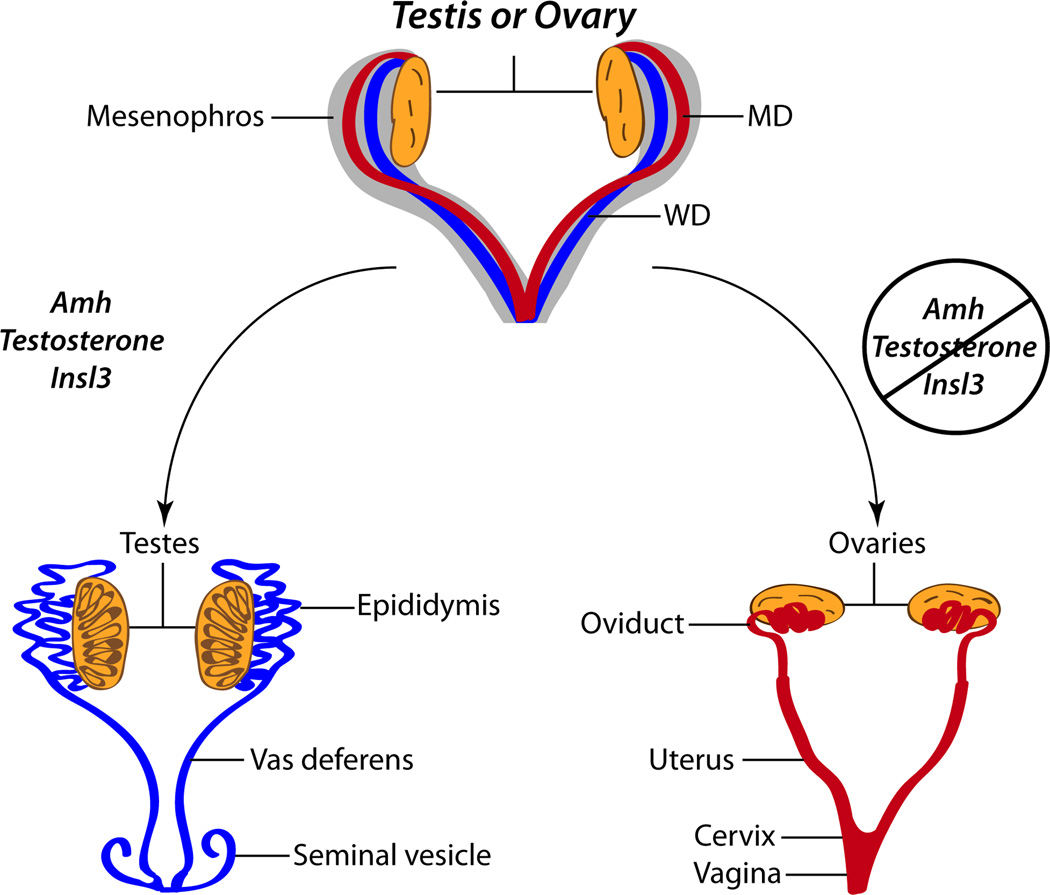
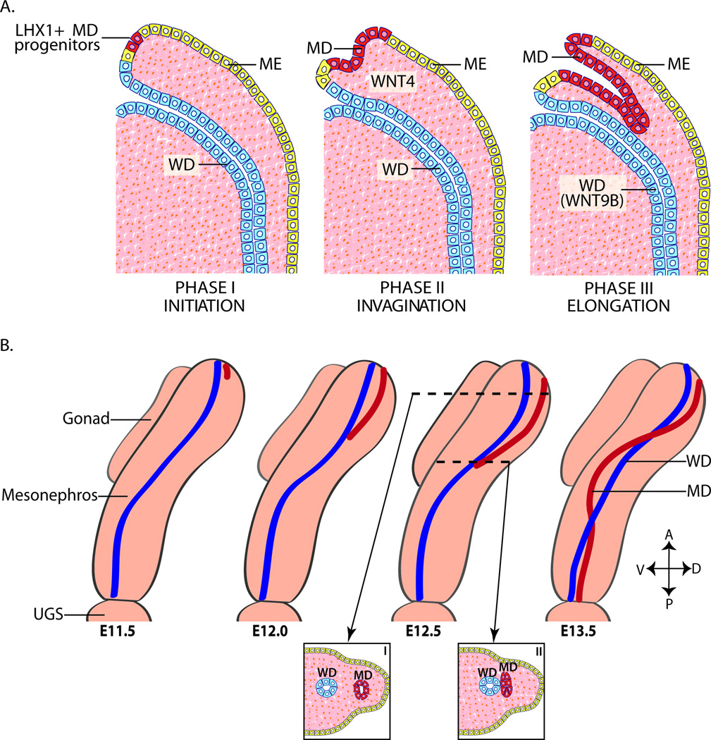
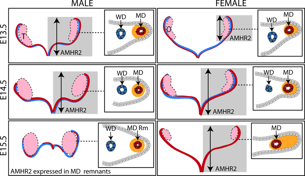
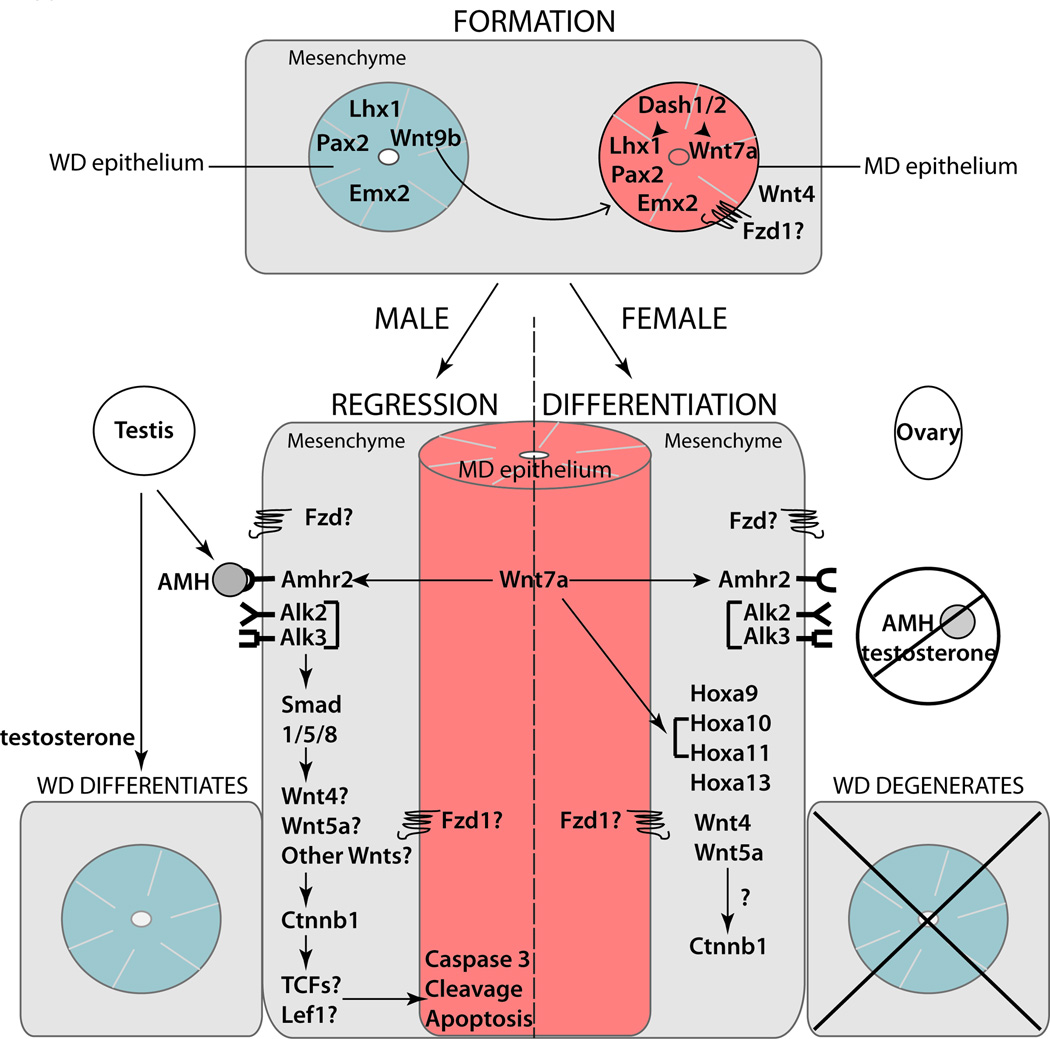
The Müllerian duct (MD) forms the female reproductive tract (FRT) consisting of the oviducts, uterus, cervix, and upper vagina. FRT function is vital to fertility, providing the site of fertilization, embryo implantation and fetal development. Developmental defects in the formation and diseases of the FRT, including cancer and endometriosis, are prevalent in humans and can result in infertility and death. Furthermore, because the MDs are initially formed regardless of genotypic sex, mesenchymal to epithelial signaling is required in males to mediate MD regression and prevents the development of MD-derived organs. In males, defects in MD regression result in the retention of FRT organs and have been described in several human syndromes. Although to date not reported in humans, ectopic activation of MD regression signaling components in females can result in aplasia of the FRT. Clearly, MD development is important to human health; however, the molecular mechanisms remain largely undetermined. Molecular genetics studies of human diseases and mouse models have provided new insights into molecular signaling during MD development, regression and differentiation. This review will provide an overview of MD development and important genes and signaling mechanisms involved.


Figures

FIG. 1
FIG. 1. Sexual differentiation of the reproductive tracts
The reproductive tracts prior to sexual differentiation are equivalent and contain a fully formed Wolffian duct (WD; represented in blue) and Müllerian duct (MD; represented in red). Hormones produced in the fetal testis, anti-Müllerian hormone (Amh), testosterone, and insulin-like 3 (Insl3), enable regression of the MD, differentiation of the WD into the male genital tract, and testicular descent, respectively. In females, lack of AMH, testosterone, and Insl3 at this developmental time permits differentiation of the MD into the female reproductive tract, passive degeneration of the WD, and maintenance of the ovaries in an abdominal position, respectively. The WD differentiates into the male reproductive tract consisting of the vas deferentia, epididymides, and seminal vesicles. The MD develops into the female reproductive tract consisting of the oviducts, uterus and upper vagina. Adapted from (Kobayashi and Behringer, 2003).
FIG. 2
FIG. 2. Müllerian duct formation
(A) MD (represented in red) formation occurs in three phases: initiation, invagination, and elongation. Phase I (Initiation); MD progenitor cells in the mesonephric epithelium (represented in yellow) are specified and begin to express LHX1. Phase II (invagination): In response to Wnt4 signaling from the mesenchyme, LHX1+ MD progenitor cells invaginate caudally into the mesenophros towards the WD (represented in blue). Phase III (Elongation): The tip of the MD contacts the WD and elongates caudally in close proximity to the WD requiring structure and WNT9B signaling from the WD. (B) Beginning at ~ E11.5 in mice, the MD invaginates and extends posteriorly guided by the WD. During elongation, mesenchymal cells separate the WD and MD anterior to the growing tip (I). However at the MD tip, the MD and WD are in contact (II). At ~E12.5, the MD crosses over the WD to be located medially. Elongation is complete by ~E13.5 with the MDs reaching the UGS. Adapted from (Kobayashi and Behringer, 2003). A, anterior (dorsal); E, embryonic day in mouse; D, dorsal; ME, mesonephric epithelium; MD, Müllerian duct; P, posterior (caudal); urogenital sinus, UGS; V, ventral; WD, Wolffian duct.
FIG. 3
FIG. 3. Müllerian duct regression
At E13.5 in mice, male and female WD (represented in blue) and MD (represented in red) are fully formed. In males, AMHR2 is expressed throughout the length (represented by gray box and arrow) and in the MD mesenchyme (represented in orange) in a tight ring around the MD epithelium (cross-section inset). In females, AMHR2 expression is caudally expressed and found in the MD mesenchyme on the antimesometrial side of the MD epithelium. By E14.5, expression of AMHR2 is found along the length of the MD in both sexes. At E14.5 in females, the WD is beginning to degenerate and is absent by E15.5. Portions of the MD have regressed in males by E15.5 in an apparently random pattern and AMHR2 expression is limited to the remaining MD remnants. E, embryonic day in mouse; MD, Müllerian duct; O, ovary; Rm, remnant; T, testis; WD, Wolffian duct.
FIG. 4
FIG. 4. Genes involved in Müllerian duct development
Multiple factors and signaling pathways in the mesenchyme (represented in gray) and epithelium of both ducts (WD epithelium represented in blue; MD epithelium represented in red) act in concert to direct MD development. Hormones and key genes required for MD formation, regression in males and differentiation in females are indicated in this figure. MD, Müllerian duct; WD, Wolffian duct.

References

    1. Allard S, Adin P, Gouedard L, di Clemente N, Josso N, Orgebin-Crist MC, Picard JY, Xavier F. Molecular mechanisms of hormone-mediated Mullerian duct regression: involvement of beta-catenin. Development. 2000;127:3349–3360. - PubMed
    1. Arango NA, Kobayashi A, Wang Y, Jamin SP, Lee HH, Orvis GD, Behringer RR. A mesenchymal perspective of Mullerian duct differentiation and regression in Amhr2-lacZ mice. Mol Reprod Dev. 2008;75:1154–1162. - PubMed
    1. Arango NA, Lovell-Badge R, Behringer RR. Targeted mutagenesis of the endogenous mouse Mis gene promoter: in vivo definition of genetic pathways of vertebrate sexual development. Cell. 1999;99:409–419. - PubMed
    1. Arango NA, Szotek PP, Manganaro TF, Oliva E, Donahoe PK, Teixeira J. Conditional deletion of beta-catenin in the mesenchyme of the developing mouse uterus results in a switch to adipogenesis in the myometrium. Dev Biol. 2005;288:276–283. - PubMed
    1. Austin HB. DiI analysis of cell migration during mullerian duct regression. Dev Biol. 1995;169:29–36. - PubMed