. 2014 Jul 29;111(30):11169-74.
doi: 10.1073/pnas.1404739111. Epub 2014 Jul 17.

Structural effects of PrP polymorphisms on intra- and interspecies prion transmission

Affiliations

Structural effects of PrP polymorphisms on intra- and interspecies prion transmission

Rachel Angers et al. Proc Natl Acad Sci U S A. .

Abstract

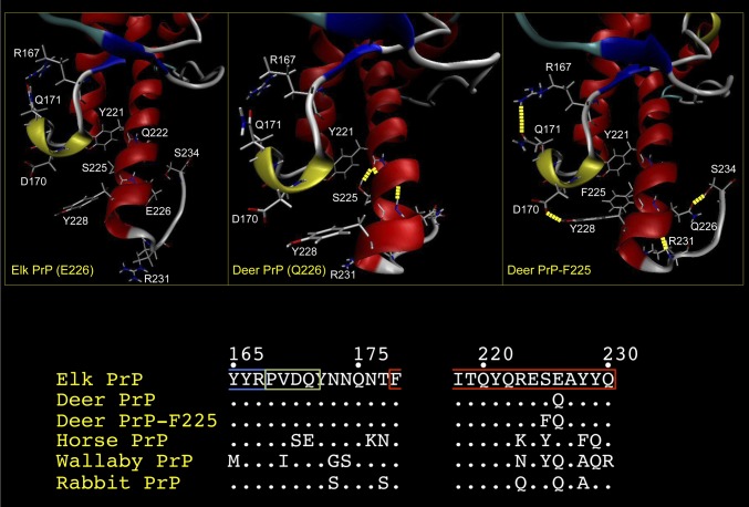
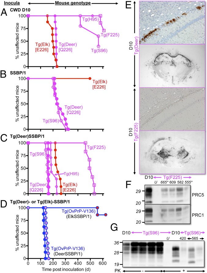
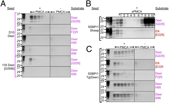
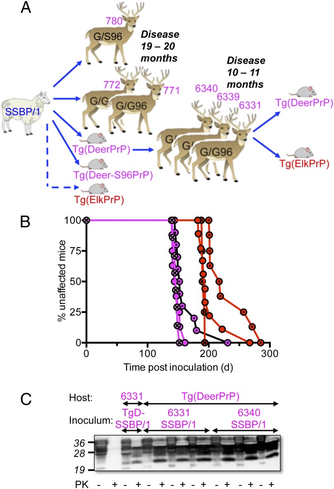
Understanding the molecular parameters governing prion propagation is crucial for controlling these lethal, proteinaceous, and infectious neurodegenerative diseases. To explore the effects of prion protein (PrP) sequence and structural variations on intra- and interspecies transmission, we integrated studies in deer, a species naturally susceptible to chronic wasting disease (CWD), a burgeoning, contagious epidemic of uncertain origin and zoonotic potential, with structural and transgenic (Tg) mouse modeling and cell-free prion amplification. CWD properties were faithfully maintained in deer following passage through Tg mice expressing cognate PrP, and the influences of naturally occurring PrP polymorphisms on CWD susceptibility were accurately reproduced in Tg mice or cell-free systems. Although Tg mice also recapitulated susceptibility of deer to sheep prions, polymorphisms that provided protection against CWD had distinct and varied influences. Whereas substitutions at residues 95 and 96 in the unstructured region affected CWD propagation, their protective effects were overridden during replication of sheep prions in Tg mice and, in the case of residue 96, deer. The inhibitory effects on sheep prions of glutamate at residue 226 in elk PrP, compared with glutamine in deer PrP, and the protective effects of the phenylalanine for serine substitution at the adjacent residue 225, coincided with structural rearrangements in the globular domain affecting interaction between α-helix 3 and the loop between β2 and α-helix 2. These structure-function analyses are consistent with previous structural investigations and confirm a role for plasticity of this tertiary structural epitope in the control of PrP conversion and strain propagation.

Keywords: prion replication; protective polymorphisms; protein structure; structural plasticity.


Conflict of interest statement

The authors declare no conflict of interest.

Figures

Fig. 1.
Fig. 1.
Structural comparisons of elk PrP, deer PrP, and deer PrP-F225. Average conformations showing both the secondary structure and the backbone structure of elk PrP (E226), deer PrP (Q226), and deer PrP-F225 obtained from 5-ns MD trajectories. Images of structures were generated using VMD. The figure focuses on the region of the protein encompassing the β2–α2 loop and α-helix 3. α-Helices are colored red; 310 helices, apple green; β-sheets, blue; turns, cyan; and coils, white. Hydrogen bonds are indicated as dashed yellow lines. Amino acid residues involved in structural differences between the three PrPs, or that vary between them, are shown in white. Only the side chains for these residues are shown. (Lower) The primary structures in the region of the β2–α2 loop and α-helix 3 of elk, deer, and deer-F225 as well as PrP from species analyzed in previous structural studies are shown.
Fig. 2.
Fig. 2.
Transgenic modeling of cervid PrP polymorphisms. (AD) Survival curves of various inoculated Tg mice. (D) Tg(OvPrP-V136) mice expressing sheep PrP with V at residue 136. Red filled circles, SSBP/1 passaged in Tg(ElkPrP); magenta filled circles, SSBP/1 passaged in Tg(DeerPrP); blue filled circles, SSBP/1. Graphs in AD have the same timescales. (E) PrPSc deposition in the region of the hippocampus and thalamus of D10-infected Tg(Deer) and Tg(F225) mice assessed by immunohistochemistry and histoblotting. (F and G) Western blots showing PrPSc in brains of diseased D10-infected Tg(F225) and Tg(S96) mice. Numbers above lanes in F and G refer to days post inoculation when mice were killed. Asterisks indicate asymptomatic mice. U, uninfected mice. In F and G, “D10” indicates D10 CWD brain. Western blots were probed with mAbs PRC5 and PRC1 as indicated.
Fig. 3.
Fig. 3.
Effects of cervid PrP substitutions on PMCA of CWD and SSBP/1. (A and C) PMCA using PrPC substrates from Tg mice. (A) Brain extracts from D10 deer or G/S96 deer 135 were used as seeds. (C) SSBP/1 adapted in Tg(DeerPrP) mice was used as seed. “+PMCA” and “–PMCA” indicate whether or not samples were subject to sonication. The first two lanes of the +PMCA panels are brain homogenates from uninfected Tg mice, untreated or treated with PK, respectively. All other samples were PK-treated. (B) Serial PMCA using PrPC from Tg(DeerPrP) or Tg(ElkPrP) mice seeded with sheep SSBP/1 prions. Numbers refer to rounds of serial PMCA. (Upper and Lower) The left four lanes contain undigested substrate; undigested SSBP/1; PK-treated SSBP/1; and unsonicated material from round 1. All other samples were PK-treated. Blots were probed with mAb 6H4.
Fig. 4.
Fig. 4.
Transmission of Tg mouse-adapted CWD prions to deer. (A) Passage history of CWD from D10 CWD deer to Tg(DeerPrP)1536+/− mice and thence to hand-raised white-tailed deer. (B) Western blots showing levels of total PrP and PrPSc in obex and RPLN of autopsied deer at 18 mo. Fifty micrograms of total protein from obex of 139 and 142 was digested; for 135, the amount was 500 μg. Western blots were probed with mAb 6H4. (C) PrPSc deposition patterns in obex assessed by immunohistochemistry. Naive, uninfected deer; CWD, deer infected with naturally occurring CWD; 142 G/G96, deer infected with Tg(DeerPrP)-passaged D10 CWD. (D) PrPSc deposition patterns in tonsil of deer 135 and 139 assessed by IHC. (Insets) Higher magnifications of individual follicles. (Scale bars in C and D, 200 μM.) (E) Survival curves of Tg(DeerPrP)1536+/− mice infected with obex (solid lines) or RPLN (dashed lines). Solid symbols, WT infected deer; open symbols, G/S96 deer. (F) PrPSc deposition patterns assessed by histoblotting in the hippocampus/thalamus of Tg(DeerPrP)1536+/− mice infected with brain homogenates from 135 and 142 deer. Blots were treated with PK as indicated.
Fig. 5.
Fig. 5.
Transmisssion of SSBP/1 to deer. (A) Passage history of SSBP/1 prions from sheep to deer and Tg mice. (B) Brain extracts from deer 6331 (circles), 6339 (circles with dots), and 6340 (circles with crosses) infected with SSBP/1 prions from Tg(DeerPrP) mice were transmitted to Tg(DeerPrP) (magenta) or Tg(ElkPrP) (red) mice. (C) Western blots of representative transmissions of SSBP/1 to deer and Tg mice.

References

    1. Will RG, et al. A new variant of Creutzfeldt-Jakob disease in the UK. Lancet. 1996;347(9006):921–925. - PubMed
    1. Prusiner SB, et al. Transgenetic studies implicate interactions between homologous PrP isoforms in scrapie prion replication. Cell. 1990;63(4):673–686. - PubMed
    1. Bessen RA, Marsh RF. Distinct PrP properties suggest the molecular basis of strain variation in transmissible mink encephalopathy. J Virol. 1994;68(12):7859–7868. - PMC - PubMed
    1. Telling GC, et al. Evidence for the conformation of the pathologic isoform of the prion protein enciphering and propagating prion diversity. Science. 1996;274(5295):2079–2082. - PubMed
    1. Angers RC, et al. Prion strain mutation determined by prion protein conformational compatibility and primary structure. Science. 2010;328(5982):1154–1158. - PMC - PubMed

Publication types

LinkOut - more resources