. 2014 Sep 5;289(36):25021-30.
doi: 10.1074/jbc.M114.565671. Epub 2014 Jul 17.

Requirements for pseudosubstrate arginine residues during autoinhibition and phosphatidylinositol 3,4,5-(PO₄)₃-dependent activation of atypical PKC

Affiliations

Requirements for pseudosubstrate arginine residues during autoinhibition and phosphatidylinositol 3,4,5-(PO₄)₃-dependent activation of atypical PKC

Robert A Ivey et al. J Biol Chem. .

Abstract

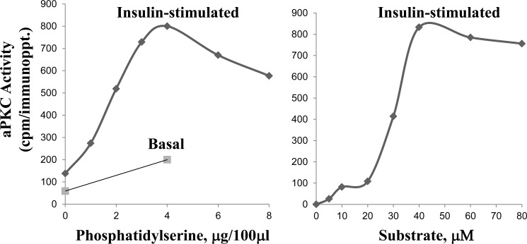
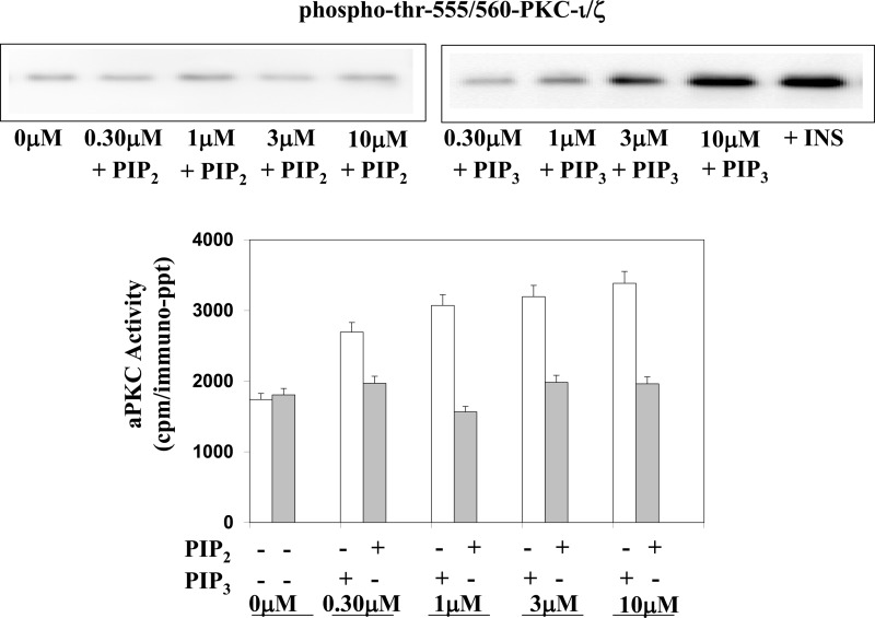
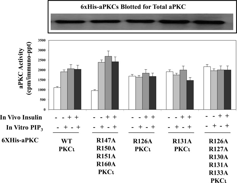
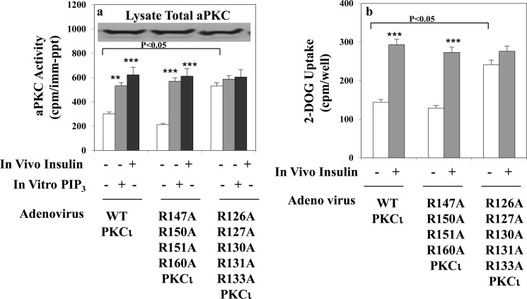
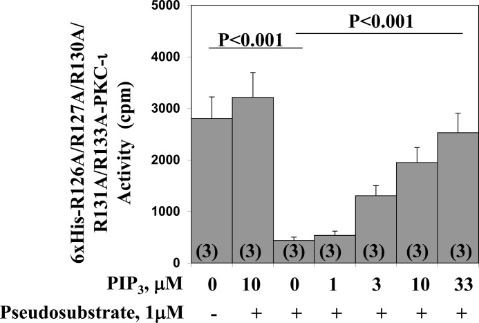
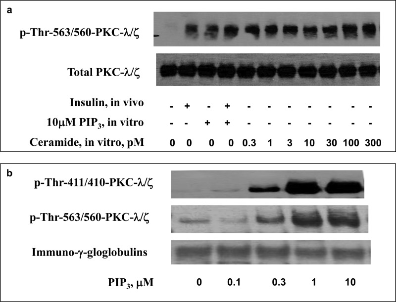
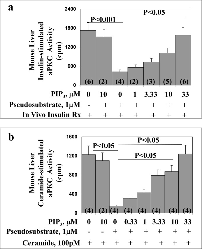
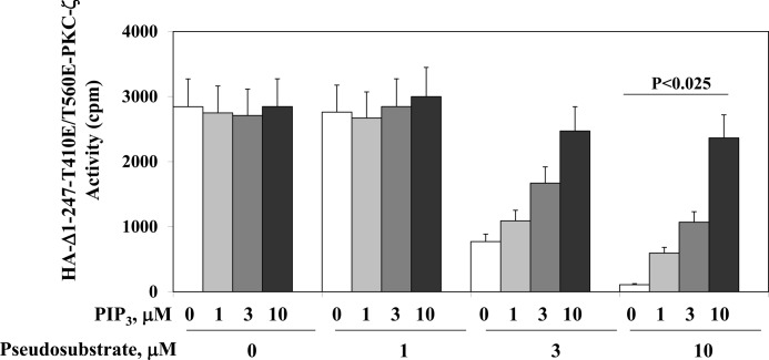
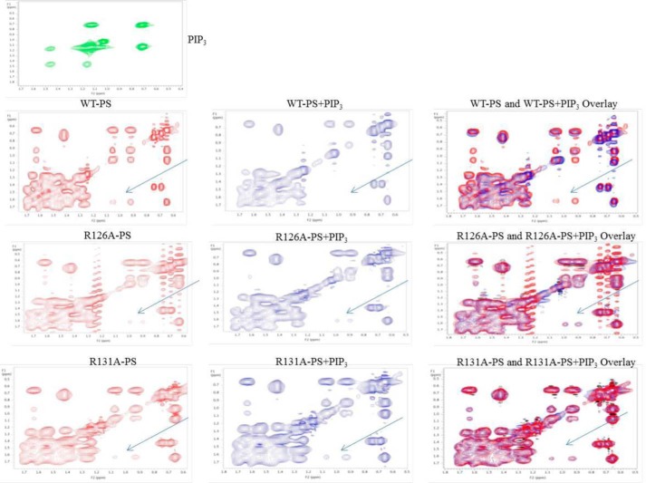
Atypical PKC (aPKC) isoforms are activated by the phosphatidylinositol 3-kinase product phosphatidylinositol 3,4,5-(PO4)3 (PIP3). How PIP3 activates aPKC is unknown. Although Akt activation involves PIP3 binding to basic residues in the Akt pleckstrin homology domain, aPKCs lack this domain. Here we examined the role of basic arginine residues common to aPKC pseudosubstrate sequences. Replacement of all five (or certain) arginine residues in the pseudosubstrate sequence of PKC-ι by site-directed mutagenesis led to constitutive activation and unresponsiveness to PIP3 in vitro or insulin in vivo. However, with the addition of the exogenous arginine-containing pseudosubstrate tridecapeptide to inhibit this constitutively active PKC-ι, PIP3-activating effects were restored. A similar restoration of responsiveness to PIP3 was seen when exogenous pseudosubstrate was used to inhibit mouse liver PKC-λ/ζ maximally activated by insulin or ceramide and a truncated, constitutively active PKC-ζ mutant lacking all regulatory domain elements and containing "activating" glutamate residues at loop and autophosphorylation sites (Δ1-247/T410E/T560E-PKC-ζ). NMR studies suggest that PIP3 binds directly to the pseudosubstrate. The ability of PIP3 to counteract the inhibitory effects of the exogenous pseudosubstrate suggests that basic residues in the pseudosubstrate sequence are required for maintaining aPKCs in an inactive state and are targeted by PIP3 for displacement from the substrate-binding site during kinase activation.

Keywords: Diabetes; Insulin Resistance; Phosphatidylinositol Kinase (PI Kinase); Protein Kinase C (PKC); Signal Transduction.


Figures

FIGURE 1.
FIGURE 1.
Dose-dependent requirements for phosphatidylserine (left panel) and substrate (right panel) during aPKC assay. Where indicated, mice were injected intraperitoneally with a maximally effective dose of insulin (1 unit/kg body weight) or saline vehicle 15 min before killing. aPKC was immunoprecipitated from liver lysates and assayed with increasing amounts of phosphatidylserine and a preferred aPKC substrate, i.e. the serine analog of the PKC-ϵ pseudosubstrate, along with other standard components of the aPKC assay (see “Experimental Procedures”). Data are mean of 2–4 replicates.
FIGURE 2.
FIGURE 2.
Effects of PIP3 and PIP2 on activity and phosphorylation of aPKC. aPKC was immunoprecipitated from lysates of livers harvested from basal (i.e. not insulin-stimulated) mice, and assayed in the presence of increasing concentrations of PIP3 (white columns) or PIP2 (black columns). Data are mean ± S.E. of triplicate determinations. Also shown are representative immunoblots of alterations in autophosphorylation (phospho-thr-563/560-PKC-λ/ζ).
FIGURE 3.
FIGURE 3.
Effects of mutation of arginine residues in the pseudosubstrate sequence of PKC-ι (R126A, R127A, R130A, R131A, and R133A) versus mutation of arginine residues contained within a ring surrounding a putative diacylglycerol activation pocket of PKC-ι (R147A, R150A, R151A, and R160A) on resting/basal aPKC activity and its activation by insulin in vivo and PIP3in vitro. Plasmids encoding WT and mutated forms of His6-tagged PKC-ι were transfected into 3T3/L1 adipocytes, and, after allowing 48–72 h for expression, cells were treated for 30 min with 100 nm insulin or left untreated. After incubation, WT and mutated forms of His6-tagged PKC-ι were adsorbed from cell lysates onto Ni-NTA beads from which the His6-tagged aPKC was eluted and assayed for aPKC activity with or without maximally effective 10 μm PIP3 as indicated. Immunoblot analysis shows nearly equal expression of the wild-type and mutated forms of His6-tagged PKC-ι as recovered in eluates of Ni-NTA beads. Data are mean ± S.E. of duplicate incubations in a representative experiment. Comparable results were obtained in two separate experiments.
FIGURE 4.
FIGURE 4.
Effects of mutation of arginine residues in the pseudosubstrate sequence of PKC-ι (R126A, R127A, R130A, R131A, and R133) versus mutation of arginine residues contained within a ring surrounding a putative diacylglycerol activation pocket of PKC-ι (R147A, R150A, R151A, and R160A) on resting/basal total cellular aPKC enzyme activity and its activation by insulin in vivo or PIP3in vitro (a) and insulin-stimulated glucose transport in intact 3T3/L1 adipocytes (b). a and b, cells were infected with multiplicity of 10 adenovirus-expressing WT or the indicated mutant forms of PKC-ι, and, after allowing 72 h for expression, the cells were treated for 30 min with 100 nm insulin or left untreated. b, cells were incubated for another 5 min, during which [3H]2-deoxyglucose uptake was measured (see “Experimental Procedures”). a, following incubation, total cellular aPKC was immunoprecipitated from cell lysates with anti-PKC-λ/ζ antiserum, and aPKC activity was measured with or without the addition of maximally effective10 μm PIP3 as indicated. Data are mean ± S.E. of four determinations. As shown in the immunoblot analysis, total cellular aPKC levels were comparable in adipocytes expressing WT and mutant forms of PKC-ι. Note that 3T3/L1 adipocytes only contain mouse PKC-ι, which is 98% homologous to human PKC-ι. **, p < 0.01; ***, p < 0.001.
FIGURE 5.
FIGURE 5.
Dose-related stimulatory effects of PIP3 on pseudosubstrate-inhibited activity of constitutively active PKC-ι in which five arginine residues in the pseudosubstrate sequence were replaced by alanine residues. The constitutive mutant form of PKC-ι, His6-126A/R127A/R130A/R131A/R133-PKC-ι, was harvested from plasmid-transfected 3T3/L1 adipocytes as described in Fig. 3 and incubated with or without a maximally effective concentration of pseudosubstrate inhibitor (1 μm), the indicated concentrations of PIP3, and other components of the aPKC assay. Data are mean ± S.E. of the number of determinations shown in parentheses.
FIGURE 6.
FIGURE 6.
Activation of mouse liver aPKC by treatment with insulin in vivo and/or PIP3 and ceramide in vitro (a) and by PIP3in vitro (b). a, where indicated, mice were treated with vehicle or insulin (1 unit/kg body weight) intraperitoneally 15 min before killing. Liver lysates were obtained, and aPKC was collected by immunoprecipitation and incubated with the indicated concentrations of ceramide and PIP3 and other components of the aPKC assay. b, aPKCs were immunoprecipitated from liver lysates of basal/untreated mice and incubated with the indicated concentrations of PIP3 and other components of the aPKC assay. After incubation, reaction mixtures were subjected to Western blot analysis for the indicated phosphoproteins and proteins. Representative blots are shown.
FIGURE 7.
FIGURE 7.
Dose-related stimulatory effects of PIP3 on pseudosubstrate-inhibited activity of mouse liver aPKC activated in vivo by insulin (panel a) or in vitro by ceramide (panel b). In panel a, liver was obtained from mice treated in vivo with insulin (1units/kg body weight) given intraperitoneally 15 min before killing. Rx, treatment. In panel b, liver was obtained from basal/untreated mice and activated during subsequent in vitro assay by addition of maximally effective 100 pm ceramide-16:0. In both cases, total aPKC was immunoprecipitated and assayed in the presence of maximally effective 1 μm pseudosubstrate and indicated concentrations of PIP3. Values are mean ± S.E. of the number of determinations shown in parentheses.
FIGURE 8.
FIGURE 8.
Dose-related stimulatory effects of PIP3 on pseudosubstrate-inhibited activity of a truncated, glutamate-pseudophosphorylated, constitutively active PKC-ζ. 3T3/L1 adipocytes were grown for 72 h with a plasmid encoding HA-tagged Δ1–247-T410E/T560E-PKC-ζ, and then the PKC-ζ mutant was harvested with anti-HA antiserum and incubated with increasing concentrations of pseudosubstrate to diminish activity of the constitutive Δ1–247-T410E/T560E-PKC-ζ mutant and increasing concentrations of PIP3 to counteract the inhibitory effects of the pseudosubstrate. Data are mean ± S.E. of two to three determinations.
FIGURE 9.
FIGURE 9.
Shown are the effects of PIP3 on the NMR spectra of WT pseudosubstrate (PS) peptide (SIYRRGARRWRKLYCANG) and arginine-replaced pseudosubstrate peptide sequences (SIYARGARRWRKLYCANG and SIYRRGARAWRKLYCANG); i.e. alanine substituted for arginine at Arg-126 and Arg-131 (R126A and R131A). Peptides were analyzed without (left column) or without PIP3 (center column). The right column shows merged spectra. Also shown is the NMR spectrum of PIP3.

References

    1. Fields A. P., Frederick L. A., Regala R. P. (2007) Targeting the oncogenic protein kinase C ι for the treatment of cancer. Biochem. Soc. Trans. 23, 1996–2000 - PubMed
    1. Beeson M., Sajan M. P., Dizon M., Grebenev D., Gomez-Daspet J., Miura A., Kanoh Y., Powe J., Bandyopadhyay G., Standaert M. L., Farese R. V. (2003) Activation of protein kinase C-ζ by insulin and phosphatidylinositol-3,4,5-(PO4)3 is defective in muscle in type 2 diabetes and impaired glucose tolerance: amelioration by rosiglitazone and exercise. Diabetes 52, 1926–1934 - PubMed
    1. Kim Y.-B., Kotani K., Ciaraldi T. P., Henry R. R., Kahn B. B. (2003) Insulin-stimulated protein kinase C-λ/ζ activity is reduced in skeletal muscle of humans with obesity and type 2 diabetes: reversal with weight reduction. Diabetes 52, 1935–1942 - PubMed
    1. Sajan M. P., Standaert M. L., Miura A., Bandyopadhyay G., Vollenweider P., Franklin D. M, Lea-Currie R., Farese R. V. (2004) Impaired activation of protein kinase C-ζ by insulin and phosphatidylinositol-3,4,5-(PO4)3 in cultured pre-adipocyte-derived adipocytes and myotubes of obese subjects. J. Clin. Endocrinol. Metab. 89, 3994–3998 - PubMed
    1. Sajan M. P., Farese R. V. (2012) Insulin signalling in hepatocytes of type 2 diabetic humans: excessive expression and activity of PKC-ι and dependent processes and reversal by PKC-ι inhibitors. Diabetologia 55, 1446–1457 - PMC - PubMed

Publication types

MeSH terms