. 2014 Jul 18;345(6194):337-41.
doi: 10.1126/science.1254198. Epub 2014 Jun 19.

Activation of cytoplasmic dynein motility by dynactin-cargo adapter complexes

Affiliations

Activation of cytoplasmic dynein motility by dynactin-cargo adapter complexes

Richard J McKenney et al. Science. .

Abstract

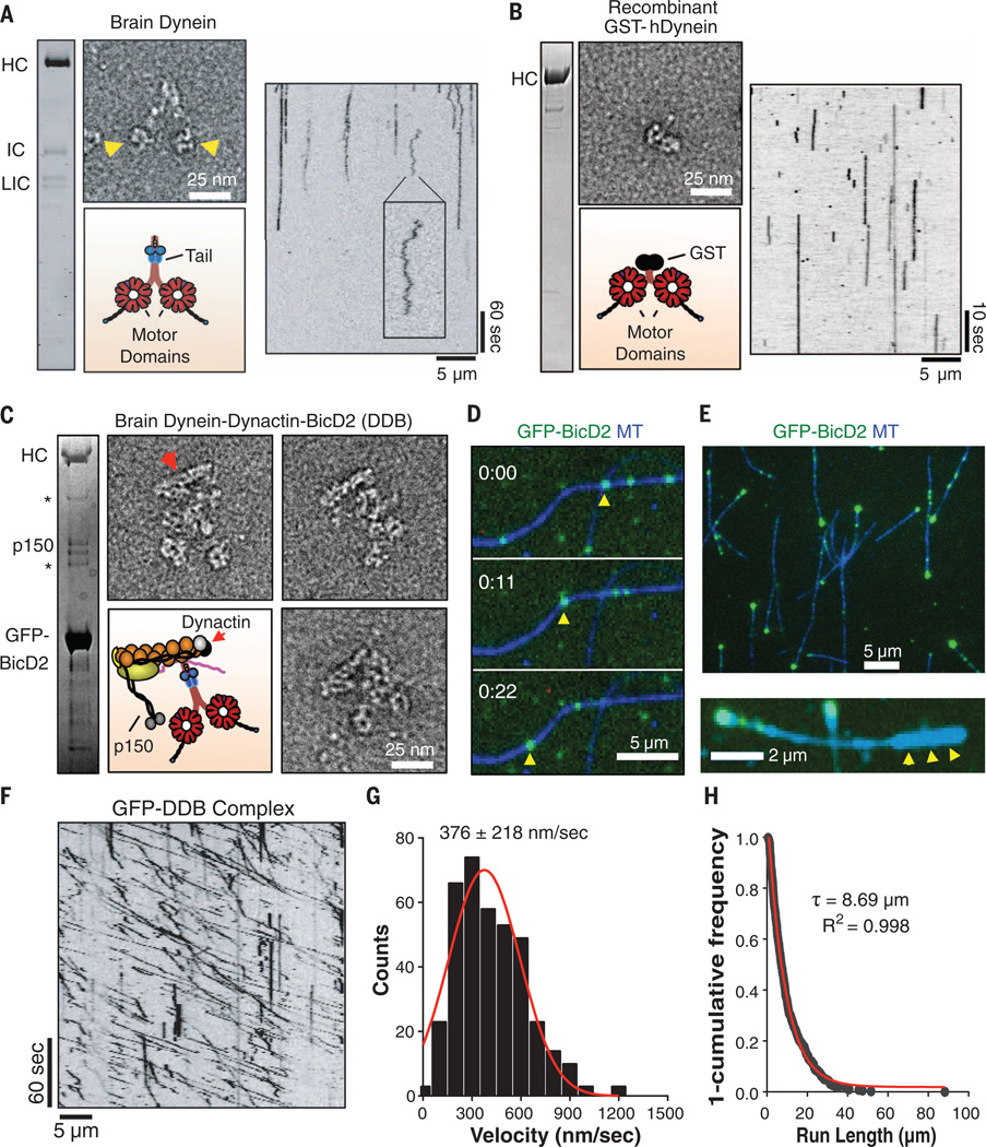
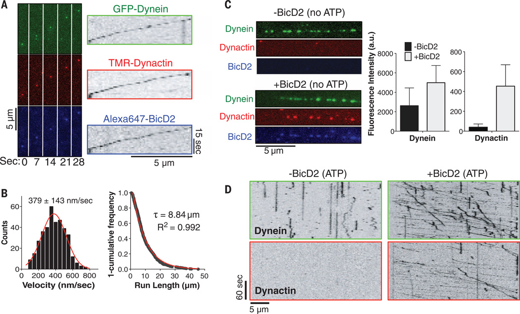
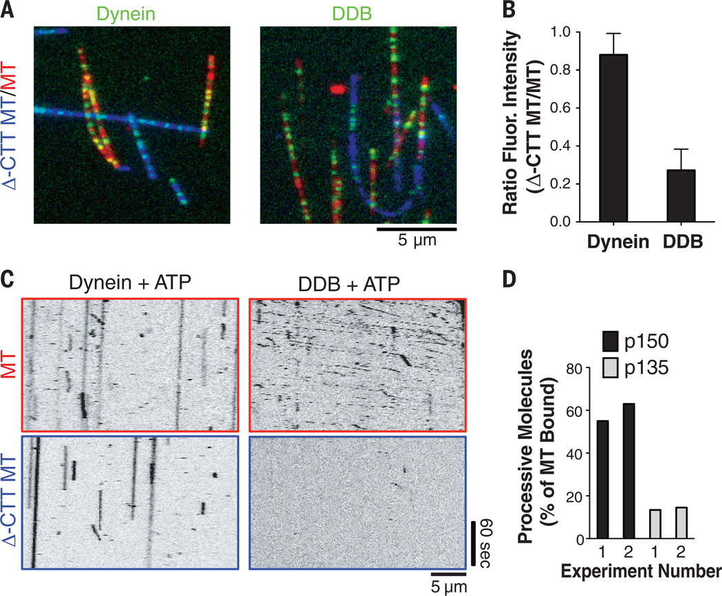
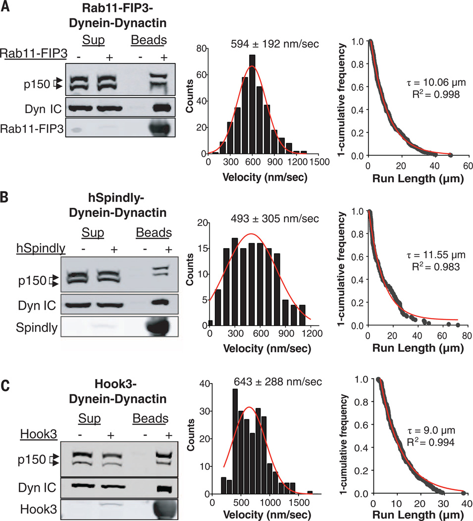
Cytoplasmic dynein is a molecular motor that transports a large variety of cargoes (e.g., organelles, messenger RNAs, and viruses) along microtubules over long intracellular distances. The dynactin protein complex is important for dynein activity in vivo, but its precise role has been unclear. Here, we found that purified mammalian dynein did not move processively on microtubules in vitro. However, when dynein formed a complex with dynactin and one of four different cargo-specific adapter proteins, the motor became ultraprocessive, moving for distances similar to those of native cargoes in living cells. Thus, we propose that dynein is largely inactive in the cytoplasm and that a variety of adapter proteins activate processive motility by linking dynactin to dynein only when the motor is bound to its proper cargo.


Figures

Fig. 1
Fig. 1. Formation of a dynein-dynactin-BicD2 complex induces processive dynein motility
(A) SDS–polyacrylamide gel electrophoresis (SDS-PAGE) of purified rat brain dynein and negative-stain EM. Arrowheads denote motor domains. Kymograph analysis of Cy3-labeled rat brain dynein on MTs (100 µM ATP). Inset: back-and-forth motion, which is likely diffusion-driven. HC: heavy chain, LIC: light intermediate chain. (B) SDS-PAGE of recombinant human GST-dynein and negative-stain EM. Kymograph analysis of TMR-labeled GST-dynein on MTs revealing no movement (100 µM ATP). (C) SDS-PAGE and negative-stain EM of purified pig brain DDB complex. Asterisks mark nonspecific bands. Red arrowhead denotes dynactin’s Arp1 filament. (D) Movie frames showing a single GFP-labeled DDB complex (yellow arrow-head) moving processively along a MT. (E) DDB complexes accumulate at one end of MTs. Below: polarity-marked MTs (arrowheads mark brightly labeled plus-end) reveal that accumulation occurs at the minus-end. (F) Kymograph analysis of pig brain DDB reveals diagonal lines reflecting long, unidirectional movements (100 µM ATP). (G) Velocity histogram(1 mM ATP) with a Gaussian fit (mean ± SD: 376 ± 218 nm/s, n = 379 molecules). (H) A “1-cumulative frequency distribution plot” run-lengths with fit to a one-phase exponential decay (red). Decay constant (τ, run length) and R2 value of the fit are shown (n = 379molecules, two independent preparations).
Fig. 2
Fig. 2. Three-color single-molecule analysis of the DDB complex and requirement of BicD2 for motility
(A) Successive frames from movie S5 showing a processive DDB complex purified from human RPE-1 cells with all three components fluorescently labeled (see text and fig S4A). Right: corresponding kymographs. (B) Histogram of human DDB velocities with a Gaussian fit (mean ± SD; n = 374 molecules). Right: a “1-cumulative frequency distribution plot” of human DDB run-lengths fit to a one-phase exponential decay (red). Decay constant (τ) and R2 value of the fit are shown (n =374 molecules). (C) Binding of fluorescently labeled human dynein and dynactin without ATP to MTs (not visible) in the absence and presence of BicD2. Note that not all DDBs are triple-labeled. Right: quantification of fluorescence along MTs (mean ± SD, n = 16 and 11 MTs before and after BicD2 addition, respectively). (D) Kymograph analysis in the presence of 100 µM ATP shows that BicD2 addition is required for human dynein motility and dynactin binding to the MTs. Note that not all dynactins in the DDB particles (+BicD2) are labeled, owing to incomplete Halo tag labeling and/or photobleaching.
Fig. 3
Fig. 3. MT binding and processivity of DDB requires the C-terminal tails of tubulin
(A) MT-binding behavior of GFP-tagged dynein (no BicD2), or GFP-DDB from human RPE cells, on normal MTs (red) and Δ-CTT MTs (blue) in the absence of ATP. (B) Quantification of the fluorescence intensity ratios (GFP-Dyn: 24 MTs, 22 Δ-CTT MTs; DDB: 59 MTs, 38 Δ-CTT MTs, mean ± SD). (C) Kymograph analysis of MT binding and motility with ATP (2 mM) on normal MTs (upper row) and on Δ-CTT MTs (lower row). (D)Quantification of processively moving (>2 µm) human DDB complexes with incorporated TMR-labeled p150-Halo or p135-Halo subunits (22) (percent of total MT bound; others were statically bound or diffusing). Results from two independent experiments are shown.
Fig. 4
Fig. 4. Rab11-FIP3, hSpindly, and Hook3 activate brain dynein motility by linking together dynein and dynactin
(A) Recombinant full-length Rab11-FIP3, (B) full-length human Spindly, and (C) human N-terminal Hook3 (amino acids 1 to 552) were prepared with an N-terminal SNAPf tag for TMR labeling (22). Left panels: All three adapters attached to beads specifically bound to dynein and dynactin from pig brain lysate (immunoblots show dynein intermediate chain and dynactin p150). Middle panels: Velocity histograms of the indicated brain dynein-dynactin-adapter complexes [Gaussian fits (red) and mean ± SD velocities are shown]. Right panels: “1-cumulative frequency distribution plot” of run-lengths for the indicated dynein-dynactin-adapter complexes. One-phase exponential decay (red), decay constants (τ) to give the run-length, and R2 values of fit are shown [n values for both middle and right panels: Rab11-FIP3 (333); hSpindly (129); Hook3 (219)].

Comment in

References

    1. Allan VJ. Biochem. Soc. Trans. 2011;39:1169–1178. - PubMed
    1. Grigoriev I, et al. Dev. Cell. 2007;13:305–314. - PubMed
    1. Griffis ER, Stuurman N, Vale RD. J. Cell Biol. 2007;177:1005–1015. - PMC - PubMed
    1. Horgan CP, Hanscom SR, Jolly RS, Futter CE, McCaffrey MW. J. Cell Sci. 2010;123:181–191. - PubMed
    1. Vallee RB, McKenney RJ, Ori-McKenney KM. Nat. Cell Biol. 2012;14:224–230. - PubMed

Publication types

Substances