Case Reports
. 2014 May;11(2):e13933.
doi: 10.5812/iranjradiol.13933. Epub 2014 May 15.

A rare cause of chronic cough: intrathoracic gossypiboma

Affiliations
Case Reports

A rare cause of chronic cough: intrathoracic gossypiboma

Dilber Yilmaz Durmaz et al. Iran J Radiol. 2014 May.

Abstract

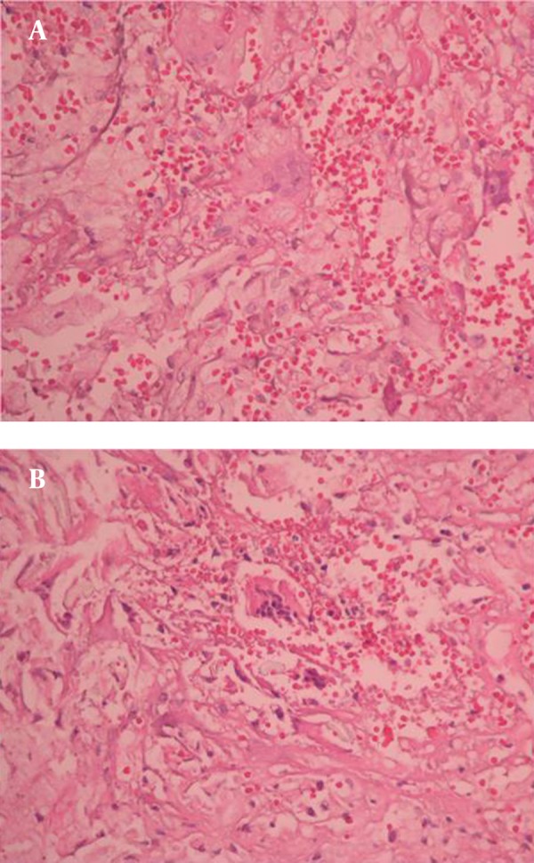
Intrathoracic gossypiboma, a retained surgical sponge in the thoracic cavity, is a rare but serious complication of thoracic surgeries. A 70-year-old man presented with an eight-month history of cough. He had undergone coronary artery bypass surgery eight years ago. The posteroanterior chest X-ray revealed a well-marginated homogeneous opacity at the left hemithorax with striped appearance in the center. Thoracic CT revealed a pleural-based mass at the left lower lobe with a hyperdense rim. After the diagnosis of gossypiboma, it was removed surgically. Although rare after thoracic surgery, gossypibomas need to be considered in the differential diagnosis in case of respiratory symptoms.

Keywords: Chronic; Cough; Thoracic Surgery.


Figures

Figure 1.
Figure 1.
A) Posteroanterior and B) Lateral chest radiograph revealed a well-marginated homogeneous opacity in the middle-lower zone of the left hemithorax with striped appearance in the center
Figure 2.
Figure 2.
A) Parenchymal and B) Mediastinal windows of thorax CT scan revealed a pleural-based mass with a hyperdense rim capsule in the laterobasal segment of the left lower lobe with hyperdense striped appearance in the center
Figure 3.
Figure 3.
A and B) The removed mass contained strong smelling purulent material and a disintegrated surgical sponge
Figure 4.
Figure 4.
Histiocyte clusters with wide and thin dense cytoplasms in fibrous stroma (H&E, 10˟)
Figure 5.
Figure 5.
A) Foreign-body giant cells containing eccentrically placed 3-15 nuclei and fine granular cytoplasm rarely displaying demonstrable foreign material (H&E, 40˟); B) Foreign-body giant cells widely displaying demonstrable foreign material (H&E, 40˟)

References

    1. Suwatanapongched T, Boonkasem S, Sathianpitayakul E, Leelachaikul P. Intrathoracic gossypiboma: radiographic and CT findings. Br J Radiol. 2005;78(933):851–3. doi: 10.1259/bjr/61657645. - DOI - PubMed
    1. Cheng TC, Chou AS, Jeng CM, Chang PY, Lee CC. Computed tomography findings of gossypiboma. J Chin Med Assoc. 2007;70(12):565–9. doi: 10.1016/s1726-4901(08)70063-7. - DOI - PubMed
    1. Madan R, Trotman-Dickenson B, Hunsaker AR. Intrathoracic gossypiboma. AJR Am J Roentgenol. 2007;189(2):W90–1. doi: 10.2214/AJR.07.2250. - DOI - PubMed
    1. Poncelet AJ, Watremez C, Tack D, Noirhomme P. Paracardiac opacity following inferior- and middle-lobe resection for bronchogenic carcinoma: unsuspected diagnosis. Chest. 2005;128(1):439–41. doi: 10.1378/chest.128.1.439. - DOI - PubMed
    1. Karabulut N, Herek D, Kiroglu Y. CT features of intrathoracic gossypiboma (textiloma). Diagn Interv Radiol. 2011;17(2):122–4. doi: 10.4261/1305-3825.DIR.3120-09.0. - DOI - PubMed

Publication types

LinkOut - more resources