Review
. 2014 Jul 17;41(1):21-35.
doi: 10.1016/j.immuni.2014.06.013.

Origin and functions of tissue macrophages

Affiliations
Review

Origin and functions of tissue macrophages

Slava Epelman et al. Immunity. .

Abstract

Macrophages are distributed in tissues throughout the body and contribute to both homeostasis and disease. Recently, it has become evident that most adult tissue macrophages originate during embryonic development and not from circulating monocytes. Each tissue has its own composition of embryonically derived and adult-derived macrophages, but it is unclear whether macrophages of distinct origins are functionally interchangeable or have unique roles at steady state. This new understanding also prompts reconsideration of the function of circulating monocytes. Classical Ly6c(hi) monocytes patrol the extravascular space in resting organs, and Ly6c(lo) nonclassical monocytes patrol the vasculature. Inflammation triggers monocytes to differentiate into macrophages, but whether resident and newly recruited macrophages possess similar functions during inflammation is unclear. Here, we define the tools used for identifying the complex origin of tissue macrophages and discuss the relative contributions of tissue niche versus ontological origin to the regulation of macrophage functions during steady state and inflammation.


Figures

Figure 1
Figure 1. Macrophage Lineages, Ontogeny, and Contribution to Populations of Resident Tissue Macrophages
(A) Schematic depicting the origins of embryonic and adult macrophage lineages. Genetic lineage tracing using Flt3Cre and direct labeling techniques, including Csf1rCreER and Runx1CreER, allow identification of adult-monocyte-derived and yolk-sac-derived macrophages. There are no available tools for lineage tracing of fetal-liver-monocyte-derived macrophages only. (B) Contribution of macrophage lineages to populations of adult resident macrophages. HSC-derived populations include embryonic populations and no definitive evidence of yolk sac origin (embryonic), whereas HSCs (adult) have passed through a FLT3+ stage (Flt3-Cre+) and are continually replaced by circulating adult monocytes.
Figure 2
Figure 2. The Life Cycle of Monocyte Subsets and Macrophages in Resting and Inflamed Tissues
In the vasculature, classical Ly6ChiCD43 monocytes give rise to nonclassical Ly6CloCD43+ monocytes through an NR4A1-dependent manner (dashed black arrow). Nonclassical monocytes go on to patrol the intravascular endothelial cell surface to clear dying endothelial cells. Classical monocytes can exit the bloodstream and have the capacity to patrol extravascular tissues. These tissue monocytes can transport antigens to lymph nodes with minimal differentiation changes from their state in blood, although a proportion can convert to Ly6Clo monocytes. However, in the context of inflammation, classical monocytes readily differentiate to macrophages. These monocyte-derived macrophages are distinct from resident macrophages, which in many organs are embryonically derived and capable of sustaining themselves by local proliferation. Both recruited and resident macrophages share the capacity for proliferation in tissue during inflammation.
Figure 3
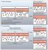
Figure 3. Schematic Representation of Repopulation Strategies Used by Tissues to Replenish Macrophage Numbers via Different Depletions Methods
(A) Cardiac macrophages. In the steady state, the majority of cardiac macrophages are embryonically derived (red, Flt3-Cre), and a smaller contribution is derived from HSCs (green, Flt3-Cre+). After depletion, Ly6chi monocytes enter into tissue, proliferate, and compete with proliferating resident cardiac macrophages to reestablish the steady-state macrophage pool. After repopulation is complete, autonomy between tissue macrophages and monocytes is restored. The pool of resident cardiac macrophages has undergone “ontological reprogramming” and is now primarily composed of adult-derived macrophages. In the absence of circulating blood monocytes (such as in Ccr2−/− mice), resident cardiac macrophages are fully capable of reestablishing steady-state levels solely through in situ proliferation. (B) Alveolar macrophages. In the steady state, the majority of alveolar macrophages are derived from embryonic progenitors, and a smaller contribution is derived from HSCs. Alveolar macrophages are instructed by GM-CSF to differentiate into functional alveolar macrophages that prevent alveolar proteinosis. After depletion (CD163-DTR), numbers of resident macrophages are reduced, and repopulation occurs solely through in situ proliferation of resident lung macrophages. If resident macrophages receive significant genotoxic injury (lethal irradiation) as a depletion strategy, recruited monocytes gain a competitive advantage and replace resident, embryonically derived lung macrophages. If recruited monocytes cannot receive GM-CSF signals, then alveolar macrophages, despite genotoxic injury, are capable of repopulating the lung macrophage niche.

References

    1. A-Gonzalez N, Guillen JA, Gallardo G, Diaz M, de la Rosa JV, Hernandez IH, Casanova-Acebes M, Lopez F, Tabraue C, Beceiro S, et al. The nuclear receptor LXRα controls the functional specialization of splenic macrophages. Nat. Immunol. 2013;14:831–839. - PMC - PubMed
    1. Ajami B, Bennett JL, Krieger C, Tetzlaff W, Rossi FM. Local self-renewal can sustain CNS microglia maintenance and function throughout adult life. Nat. Neurosci. 2007;10:1538–1543. - PubMed
    1. Ajami B, Bennett JL, Krieger C, McNagny KM, Rossi FM. Infiltrating monocytes trigger EAE progression, but do not contribute to the resident microglia pool. Nat. Neurosci. 2011;14:1142–1149. - PubMed
    1. Amano SU, Cohen JL, Vangala P, Tencerova M, Nicoloro SM, Yawe JC, Shen Y, Czech MP, Aouadi M. Local proliferation of macrophages contributes to obesity-associated adipose tissue inflammation. Cell Metab. 2014;19:162–171. - PMC - PubMed
    1. Arnold T, Betsholtz C. The importance of microglia in the development of the vasculature in the central nervous system. Vascular Cell. 2013;5:4. - PMC - PubMed