Review
. 2014 Aug;115(2-3):294-304.
doi: 10.1016/j.pbiomolbio.2014.07.001. Epub 2014 Jul 15.

Cardiac applications of optogenetics

Affiliations
Review

Cardiac applications of optogenetics

Christina M Ambrosi et al. Prog Biophys Mol Biol. 2014 Aug.

Abstract

In complex multicellular systems, such as the brain or the heart, the ability to selectively perturb and observe the response of individual components at the cellular level and with millisecond resolution in time, is essential for mechanistic understanding of function. Optogenetics uses genetic encoding of light sensitivity (by the expression of microbial opsins) to provide such capabilities for manipulation, recording, and control by light with cell specificity and high spatiotemporal resolution. As an optical approach, it is inherently scalable for remote and parallel interrogation of biological function at the tissue level; with implantable miniaturized devices, the technique is uniquely suitable for in vivo tracking of function, as illustrated by numerous applications in the brain. Its expansion into the cardiac area has been slow. Here, using examples from published research and original data, we focus on optogenetics applications to cardiac electrophysiology, specifically dealing with the ability to manipulate membrane voltage by light with implications for cardiac pacing, cardioversion, cell communication, and arrhythmia research, in general. We discuss gene and cell delivery methods of inscribing light sensitivity in cardiac tissue, functionality of the light-sensitive ion channels within different types of cardiac cells, utility in probing electrical coupling between different cell types, approaches and design solutions to all-optical electrophysiology by the combination of optogenetic sensors and actuators, and specific challenges in moving towards in vivo cardiac optogenetics.

Keywords: Cardiomyocytes; Cell delivery; Channelrhodopsin; Fibroblasts; Gene delivery; Optogenetics.


Figures

Figure 1
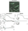
Figure 1. ChR2 current during the cardiac action potential via AP clamp
A. Adult guinea pig ventricular cardiomyocytes after 48h of viral infection with Ad-ChR2(H134R)-EYFP, green fluorescence indicates ChR2 expression; scale bar is 50 μm. Experimental (B–D) and modeling (E–F) traces for guinea pig ventricular cells. B. Optically-triggered action potential (50 ms pulse at 470nm, 1.5 mW/mm2) used for the AP clamp; dotted line indicates the voltage clamp conditions upon application of the waveform; blue dots indicate the beginning and end of the optical pulse. C. Extracted IChR2 as the difference current from the total current traces (panel D) recorded in dark conditions and with a light pulse. E. Analogous optically-triggered action potential in a model of a guinea pig ventricular cell. F. The underlying IChR2 according to the model. Reproduced with permission from (Williams et al., 2013).
Figure 2
Figure 2. Optical excitability of cardiac syncytium in vitro as a function of delivery mode
A,B. Binarized images of transgene distributions resulting from adenoviral delivery of ChR2-eYFP (ChR2 density, 54%) and cell delivery via ChR2-eYFP-expressing HEK cells (ChR2 density, 59%). White pixels represent cells expressing ChR2-eYFP and black pixels represent unmodified cells. Scale bars are 1 mm. C. Optical excitation thresholds at 50 ms pulse width for both modes of delivery. Data presented as mean ± SEM.
Figure 3
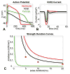
Figure 3. Optical excitation in human cardiac cell types
A. Optically-triggered action potentials (10 ms pulse at 470nm, 0.5 mW/mm2) in human ventricular, atrial and Purkinje cells. B. Underlying ChR2 current upon the action potential generation for the three cell types. C. Strength-duration curves for the three cell types. Squares show simulated values for optical excitation threshold in ventricular myocytes with a Purkinje formulation of IK1. Reproduced with permission from (Williams et al., 2013).
Figure 4
Figure 4. ChR2 expression in cardiomyocytes and cardiac fibroblasts
A. ChR2-eYFP expression localizes to the cell membrane in cardiomyocytes. B. ChR2-eYFP expression is more diffuse cytoplasmic. C. Quantification of ChR2 expression patterns in cardiomyocytes and fibroblasts; inset is a summary box plot. Line inside box represents median; bars above and below box represents max and min; box represents the 25th and 75th percentile. Scale bars are 50 μm.
Figure 5
Figure 5
A. Activation spectra for ChR2, and excitation (EX) and emission (EM) spectra for the voltage sensitive dye Di-4-ANBDQPQ and the calcium sensitive dye Rhod-4. B. Conceptual schematic of the optical path for all-optical interrogation.
Figure 6
Figure 6
All-optical measurements in cardiomyocytes using ChR2 as actuator and Rhod-4 as a sensor – example traces, comparing: A. electrical pacing and B. optical pacing of ChR2 expressing neonatal cardiomyocytes. Top: Rhod-4 signal (Ca2+); Bottom: Contractility measurements obtained through videotracking simultaneously with the Ca2+ data.
Figure 7
Figure 7. In vitro and in vivo cardiac expression of AAV1
A. Neonatal rat ventricular cardiomyocytes infected with AAV1 in vitro at MOI 1000. Green fluorescence indicates eGFP reporter and blue indicates DAPI nuclear staining. Scale bar is 50 μm. B. Schematic of a slice of an adult rat heart infected with 1×10¹¹ AAV1 particles, delivered via intramyocardial injection 4 weeks earlier, as indicated by the arrow.

References

    1. Abilez OJ, Wong J, Prakash R, Deisseroth K, Zarins CK, Kuhl E. Multiscale computational models for optogenetic control of cardiac function. Biophys J. 2011;101:1326–34. - PMC - PubMed
    1. Acharya A, Baek ST, Banfi S, Eskiocak B, Tallquist MD. Efficient inducible Cre-mediated recombination in Tcf21 cell lineages in the heart and kidney. Genesis. 2011;49:870–7. - PMC - PubMed
    1. Addis RC, Ifkovits JL, Pinto F, Kellam LD, Esteso P, Rentschler S, Christoforou N, Epstein JA, Gearhart JD. Optimization of direct fibroblast reprogramming to cardiomyocytes using calcium activity as a functional measure of success. J Mol Cell Cardiol. 2013;60:97–106. - PMC - PubMed
    1. Airan RD, Thompson KR, Fenno LE, Bernstein H, Deisseroth K. Temporally precise in vivo control of intracellular signalling. Nature. 2009;458:1025–9. - PubMed
    1. Ambrosi CM, Chen K, Kalra J, Chen X, Zhu W, Yu J, Jiang YP, Lin RZ, Cohen IS, Entcheva E. Book Distribution of donor cells alters energy requirements for optogenetic control of the heart. City: 2013. Distribution of donor cells alters energy requirements for optogenetic control of the heart.

Publication types

MeSH terms