. 2014 Jul 18;9(7):e101879.
doi: 10.1371/journal.pone.0101879. eCollection 2014.

Tubular overexpression of gremlin induces renal damage susceptibility in mice

Affiliations

Tubular overexpression of gremlin induces renal damage susceptibility in mice

Alejandra Droguett et al. PLoS One. .

Erratum in

  • PLoS One. 2014;9(10):e112003

Abstract

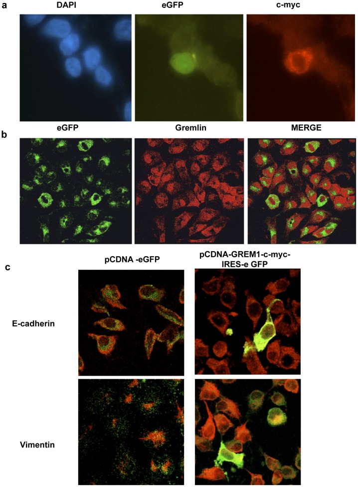
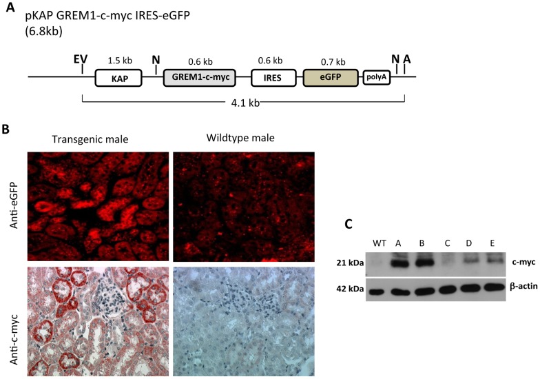
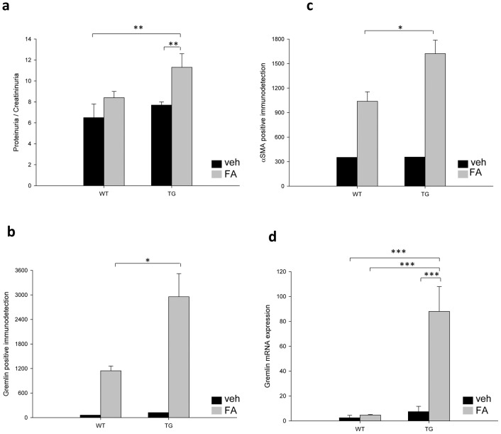
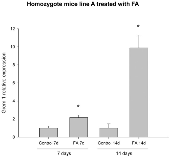
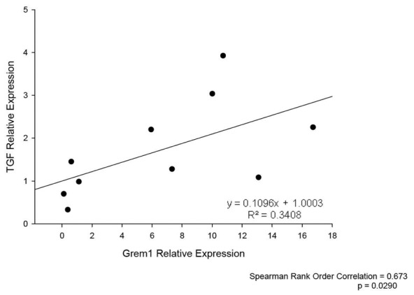
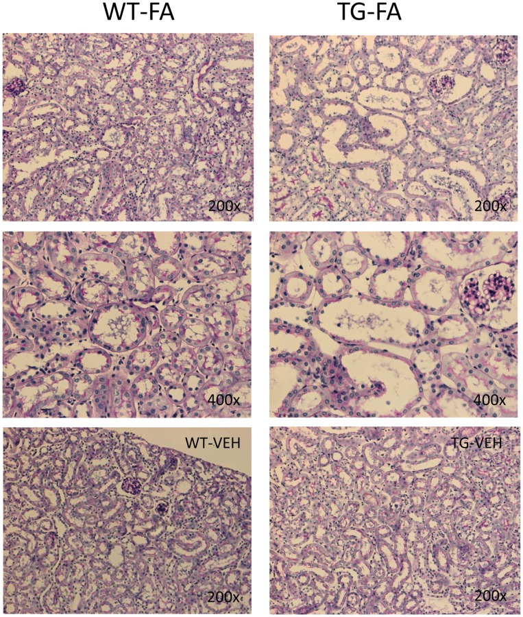
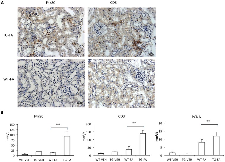
A growing number of patients are recognized worldwide to have chronic kidney disease. Glomerular and interstitial fibrosis are hallmarks of renal progression. However, fibrosis of the kidney remains an unresolved challenge, and its molecular mechanisms are still not fully understood. Gremlin is an embryogenic gene that has been shown to play a key role in nephrogenesis, and its expression is generally low in the normal adult kidney. However, gremlin expression is elevated in many human renal diseases, including diabetic nephropathy, pauci-immune glomerulonephritis and chronic allograft nephropathy. Several studies have proposed that gremlin may be involved in renal damage by acting as a downstream mediator of TGF-β. To examine the in vivo role of gremlin in kidney pathophysiology, we generated seven viable transgenic mouse lines expressing human gremlin (GREM1) specifically in renal proximal tubular epithelial cells under the control of an androgen-regulated promoter. These lines demonstrated 1.2- to 200-fold increased GREM1 expression. GREM1 transgenic mice presented a normal phenotype and were without proteinuria and renal function involvement. In response to the acute renal damage cause by folic acid nephrotoxicity, tubule-specific GREM1 transgenic mice developed increased proteinuria after 7 and 14 days compared with wild-type treated mice. At 14 days tubular lesions, such as dilatation, epithelium flattening and hyaline casts, with interstitial cell infiltration and mild fibrosis were significantly more prominent in transgenic mice than wild-type mice. Tubular GREM1 overexpression was correlated with the renal upregulation of profibrotic factors, such as TGF-β and αSMA, and with increased numbers of monocytes/macrophages and lymphocytes compared to wild-type mice. Taken together, our results suggest that GREM1-overexpressing mice have an increased susceptibility to renal damage, supporting the involvement of gremlin in renal damage progression. This transgenic mouse model could be used as a new tool for enhancing the knowledge of renal disease progression.


Conflict of interest statement

Competing Interests: The authors have declared that no competing interests exist.

Figures

Figure 1
Figure 1. In vitro validation of the GREM1 plasmid.
(a) EBNA293 cells were transfected with pCDNA3-GREM1-c-myc-IRES-eGFP, as described in the methods. Immunofluorescence shows that eGFP (green) and c-myc (red) are expressed in the same transfected EBNA293 cell. Nuclei were stained with DAPI (blue) (1000x). (b) In HK-2 cells transfected with pCDNA3-GREM1-c-myc-IRES-eGFP, GREM-1 expression was evaluated by immunocytochemistry using an antibody against GREM-1, followed by a secondary TRICT antibody (red staining). The figure shows eGFP and GREM-1 expressed in the same cell (800x). (c) Confocal immunofluorescence of HK-2 transfected cells, showing the loss of E-cadherin and induction of vimentin in eGFP-positive GREM-1-expressing cells (1600X). E-cadherin and vimentin immunostaining was detected with secondary anti-FITC antibodies (green).
Figure 2
Figure 2. Generation and validation of transgenic mice with specific tubular GREM1 overexpression.
(a) Illustration of the pKAP GREM1-c-myc-IRES-eGFP plasmid. Restriction sites used for transgene isolation are indicated with EV (EcoRV) and A (AseI). (b) eGFP and c-myc detection in renal tubular epithelial cells of transgenic mice. Immunofluorescence against eGFP and immunohistochemistry for c-myc (peroxidase immunostaining) to detect these proteins in the kidney tissue of transgenic males from line A and WT mice (400x). (c) Kidneys were dissected from WT and transgenic male mice of lines A, B, C, D and E, and isolated proteins were subjected to western blotting using an antibody against c-myc (1∶1000); anti β-actin (1∶2500) was used as a loading control. GREM1 expression was determined by densitometric analysis of the c-myc/β-actin ratio and normalized to transgenic line C expression.
Figure 3
Figure 3. FA injection induces proteinuria and GREM1 and αSMA expression in transgenic line D.
(a) The urinary protein to creatinine ratio (µg/mg) was examined in each experimental group. Positive IHC signals were quantified for (b) gremlin and (c) αSMA with KS300 image analyzer software. (d) Gremlin mRNA expression levels were determined using real-time PCR. The four parameters were significantly increased in GREM1-overexpressing transgenic mice. Data are shown as the mean ± SEM of 5-6 mice per group * p < 0.05; ** p < 0.01; *** p < 0.001. TG vs WT control
Figure 4
Figure 4. Effect of FA administration on GREM1 expression in transgenic line A homozygous mice.
Gremlin expression in transgenic mice was examined 7 and 14 days after treatment with FA. The GREM1 relative expression in homozygous mice of transgenic line A was increased at 7 days after injection with FA and remained increased at 14 days. Data are shown as the mean ± SEM of 4-9 mice per group * p≤0.0049 TG-FA vs TG-Veh, used as control because no WT littermates of the transgenic homozygotes mice were available.
Figure 5
Figure 5. Correlation of TGF-β and GREM1 expression in transgenic line A homozygous mice.
TGF-β gene expression was measured in FA-injected GREM1 transgenic mice. We observed a strongly positive correlation between TGF-β and GREM1 expression (p≤0.029, R  =  0.67; 4-9 mice per group).
Figure 6
Figure 6. Histological analysis (PAS, Masson) of FA-injected mice in transgenic line A homozygous mice.
TG-FA mice (right column) showed more severe morphological lesions (tubular dilatation, flattening of tubular epithelial cells, hyaline casts, interstitial infiltrating cells and mild interstitial fibrosis) compared with WT mice (left column). p < 0.05 (200x-400x). Control vehicle treated mice are shown at bottom. Figure shows representative mice of each group of 14–18 studied.
Figure 7
Figure 7. FA injection induces interstitial cell infiltration in transgenic line A homozygous mice.
The inflammatory cell infiltration was characterized by immunohistochemistry with anti-F4/80 (monocytes/macrophages) and anti-CD3 (T cells) antibodies. (A and B). Representative immunostaining of one mouse from each group (x400 magnification). (C) Quantification of positive IHC signals were quantified for (a) F4/80, (b) CD3 and (c) PCNA using KS300 image analyzer software. All parameters were significantly increased in transgenic mice. Data are shown as the mean ± SEM of 14–18 mice per group * p < 0.05; ** p < 0.01 vs WT-FA.

References

    1. Topol LZ, Marx M, Laugier D, Bogdanova NN, Boubnov NV, et al. (1997) Identification of dmr, a novel gene whose expression is suppressed in transformed cells and which can inhibit growth of normal but not transformed cells in culture. Mol Cell Biol 17: 4801–4810. - PMC - PubMed
    1. Topol LZ, Bardot B, Zhang Q, Resau J, Huillard E, et al. (2000) Biosynthesis, post-translation modification, and functional characterization of Drm/Gremlin. J Biol Chem 275: 8785–8793. - PubMed
    1. Hsu DR, Economides AN, Wang X, Eimon PM, Harland RM (1998) The Xenopus dorsalizing factor Gremlin identifies a novel family of secreted proteins that antagonize BMP activities. Mol Cell 1: 673–683. - PubMed
    1. Michos O, Panman L, Vintersten K, Beier K, Zeller R, et al. (2004) Gremlin-mediated BMP antagonism induces the epithelial-mesenchymal feedback signaling controlling metanephric kidney and limb organogenesis. Development 131: 3401–3410. - PubMed
    1. Roxburgh SA, Murphy M, Pollock CA, Brazil D (2006) Recapitulation of embryological programmes in renal fibrosis–the importance of epithelial cell plasticity and developmental genes. Nephron Physiol 103: 139–148. - PubMed

Publication types

Substances