. 2014 Jul 18;9(7):e102342.
doi: 10.1371/journal.pone.0102342. eCollection 2014.

Vagus nerve stimulation attenuates cerebral ischemia and reperfusion injury via endogenous cholinergic pathway in rat

Affiliations

Vagus nerve stimulation attenuates cerebral ischemia and reperfusion injury via endogenous cholinergic pathway in rat

Ying Jiang et al. PLoS One. .

Abstract

Inflammation and apoptosis play critical roles in the acute progression of ischemic injury pathology. Emerging evidence indicates that vagus nerve stimulation (VNS) following focal cerebral ischemia and reperfusion (I/R) may be neuroprotective by limiting infarct size. However, the underlying molecular mechanisms remain unclear. In this study, we investigated whether the protective effects of VNS in acute cerebral I/R injury were associated with anti-inflammatory and anti-apoptotic processes. Male Sprague-Dawley (SD) rats underwent VNS at 30 min after focal cerebral I/R surgery. Twenty-four h after reperfusion, neurological deficit scores, infarct volume, and neuronal apoptosis were evaluated. In addition, the levels of pro-inflammatory cytokines were detected using enzyme-linked immune sorbent assay (ELISA), and immunofluorescence staining for the endogenous "cholinergic anti-inflammatory pathway" was also performed. The protein expression of a7 nicotinic acetylcholine receptor (a7nAchR), phosphorylated Akt (p-Akt), and cleaved caspase 3 in ischemic penumbra were determined with Western blot analysis. I/R rats treated with VNS (I/R+VNS) had significantly better neurological deficit scores, reduced cerebral infarct volume, and decreased number of TdT mediated dUTP nick end labeling (TUNEL) positive cells. Furthermore, in the ischemic penumbra of the I/R+VNS group, the levels of pro-inflammatory cytokines and cleaved caspase 3 protein were significantly decreased, and the levels of a7nAchR and phosphorylated Akt were significantly increased relative to the I/R alone group. These results indicate that VNS is neuroprotective in acute cerebral I/R injury by suppressing inflammation and apoptosis via activation of cholinergic and a7nAchR/Akt pathways.


Conflict of interest statement

Competing Interests: The authors have declared that no competing interests exist.

Figures

Figure 1
Figure 1. The Experimental Protocol.
I/R and I/R+VNS groups underwent transient MCAO surgery. In the I/R+VNS and sham+VNS group, VNS was initiated at 30 min after occlusion and repeated every 5 min for 1 h. At 120 min, I/R and I/R+VNS groups underwent reperfusion. During the experiment, the mean CVA, HR, and blood gas were measured at the designated time points. Laser doppler flowmetry monitored the changes of right cerebral middle artery blood flow, and temperature was maintained at approximately 37°C. Neurological deficit scores assessment was performed at 3 and 24 h after reperfusion.
Figure 2
Figure 2. Time course of CVA during the experiment.
Changes in tail arterial pressure are shown for the three groups during the experiment. The time points are as follows: baseline (BS), after occlusion but before stimulation (BVNS), during the stimulation (DVNS), after stimulation but before reperfusion (AVNS), and after reperfusion (AR). * p<0.05 relative to the time point (BVNS). #p<0.05 relative to the time point (AVNS).
Figure 3
Figure 3. Time course of HR during the experiment.
HR is plotted for the same time points as in Fig. 2. HR and BP immediately fell during each pulse train and recovered to baseline values upon cessation of the stimulation in I/R+VNS and sham+VNS groups. * p<0.05 in comparison to the time point (BVNS). #p<0.05 in comparison to the time point (AVNS).
Figure 4
Figure 4. Effect of VNS on regional blood flow of right middle cerebral artery.
Changes of CBF in the right middle cerebral artery territory in three group (n = 8/group). Data are shown as mean±SEM.(P>0.05).
Figure 5
Figure 5. Neurological deficit scores after I/R with and without VNS.
Bar graphs show the neurological deficit scores at 3 and 24 h following reperfusion in the three groups. Data are shown as mean±SEM *p<0.05.
Figure 6
Figure 6. Neuroprotection induced by VNS on the infarct volume following I/R.
TTC staining in the three groups at the level of bregma 0.4mm. A: sham+VNS group, B: I/R group, and C: I/R+VNS group. D. Histograms show that the relative percentage of infarct volume 24 h after acute I/R. Red area is the healthy tissue, and the white area is the infarct tissue. *p<0.05 with relative to the I/R group, #p<0.05 relative to sham+VNS group.
Figure 7
Figure 7. Effect of VNS on neuronal apoptosis in ischemic penumbra.
Representative photomicrographs of neuron apoptosis in right ischemic penumbra cortex 24(magnification 400×). A: sham+VNS group, B: I/R group, C: I/R+VNS group. D: Quantitative analysis of the number of TUNEL-positive cells in the ischemic penumbra. Data are expressed as mean ±SEM; *P<0.05 vs I/R group, #p<0.05 vs sham+VNS group, scar = 100 µm.
Figure 8
Figure 8. Effect of VNS on inflammatory cytokines in the ischemic penumbra at 24 h after reperfusion.
The concentrations of TNF-a, IL-1β, and IL-6 of the peri-infarct region were measured by ELISA. A: TNF-a, B: IL-1β, C: IL-6 at 24 h reperfusion in the peri-infarct region. Data are presented as mean±SEM (n = 15 per group); *P<0.05 vs I/R group, #p<0.05 vs sham+VNS group.
Figure 9
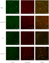
Figure 9. Immunostaining for a7nAchR on microglia (Iba-1) surface in ischemic penumbra.
The expression of a7nAchR was reduced following I/R and increased by VNS. Underwent ischemic insults, the morphology of the microglial cell changed. A: Staining for a7nAchR in red, B: Staining (microglia) Iba-1 in green, C: merge of A and B. scare bar = 75 µm.
Figure 10
Figure 10. Expression of a7nAchR, P-Akt, and cleaved caspase 3 protein in the ischemic penumbra.
Western blot showing the differential protein expression of a7nAchR, P-Akt, and cleaved caspase-3 24 h following reperfusion in the ischemic penumbra of rats among the three groups. β-actin is a loading control, showing equal loading of protein. A: sham+VNS group; B: I/R group; C: I/R+VNS group. (A) The density of a-7nAchR, (B) p-Akt, and (C) cleaved caspase 3 protein expression normalized to β-actin protein expression. *P<0.05 relative to the I/R group, #p<0.05 relative to the sham+VNS group, n = 8/group.

References

    1. Brouns R, De Deyn PP (2009) The complexity of neurobiological processes in acute ischemic stroke. Clin Neurol Neurosurg 111: 483–495. - PubMed
    1. Wang Q, Tang XN, Yenari MA (2007) The inflammatory response in stroke. J Neuroimmunol 184: 53–68. - PMC - PubMed
    1. Wang JY, Shen J, Gao Q, Ye ZG, Yang SY, et al. (2008) Ischemic postconditioning protects against global cerebral ischemia/reperfusion-induced injury in rats. Stroke 39: 983–990. - PubMed
    1. Fu SH, Zhang HF, Yang ZB, Li TB, Liu B, et al. (2014) Alda-1 reduces cerebral ischemia/reperfusion injury in rat through clearance of reactive aldehydes. Naunyn Schmiedebergs Arch Pharmacol 387: 87–94. - PubMed
    1. You SJ, Kang HC, Kim HD, Ko TS, Kim DS, et al. (2007) Vagus nerve stimulation in intractable childhood epilepsy: a Korean multicenter experience. J Korean Med Sci 22: 442–445. - PMC - PubMed

Publication types

MeSH terms