. 2014 Aug 6;136(31):10858-61.
doi: 10.1021/ja5053279. Epub 2014 Jul 24.

Hand-held and integrated single-cell pipettes

Affiliations

Hand-held and integrated single-cell pipettes

Kai Zhang et al. J Am Chem Soc. .

Abstract

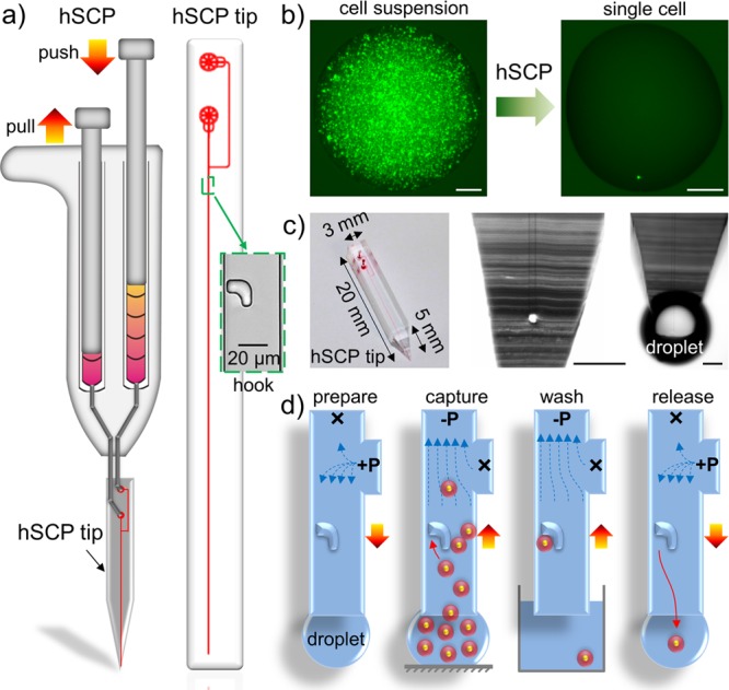
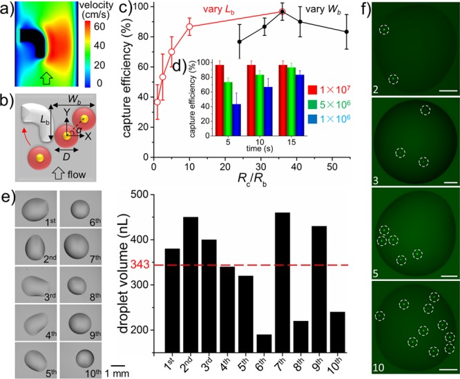
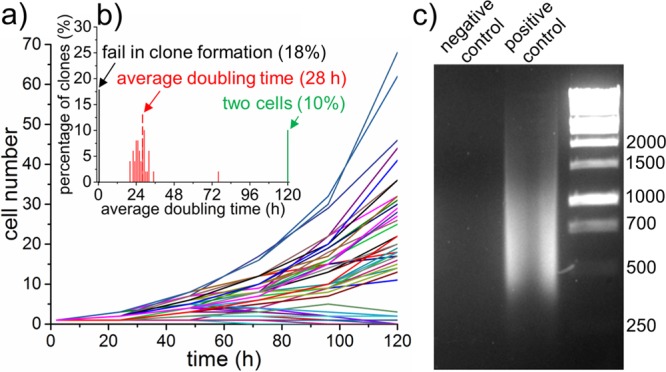
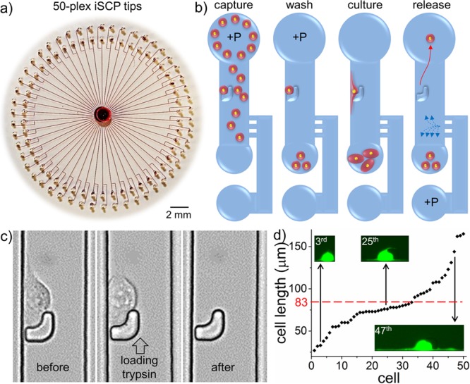
Successful single-cell isolation is a primary step for subsequent chemical and biological analyses of single cells. Conventional single-cell isolation methods often encounter operational complexity, limited efficiency, deterioration of cell viability, incompetence in the isolation of a single-cell into nanoliter liquid, and/or inability to select single adherent cells with specific phenotypes. Here, we develop a hand-held single-cell pipet (hSCP) that is rapid, operationally simple, highly efficient, and inexpensive for unbiased isolation of single viable suspended cells directly from submicroliter cell suspensions into nanoliter droplets without the assistance of any additional equipment. An integrated SCP (iSCP) has also been developed for selective isolation of single suspended and adherent cells according to the fluorescence imaging and morphological features. The isolated single cells can be conveniently transferred into standard 96-/384-well plates, Petri dishes, or vials for cloning, PCR, and other single-cell biochemical assays.


Figures

Figure 1
Figure 1
Design and mechanism of the hand-held single-cell pipet (hSCP). (a) The hSCP involves one dual-channel pipet and one hSCP tip. A magnified hook for single-cell capture is shown. (b) A single calcein-labeled SK-BR-3 cell is isolated directly from a dense cell suspension by hSCP. (c) The hSCP tip with a conical end and two magnified tip ends shown before and after extrusion of aqueous solution. (d) Work flow for single-cell isolation using hSCP. Scale bar, 200 μm.
Figure 2
Figure 2
Optimization and characterization of hSCP for single-cell manipulation. (a) Numerical simulation of flow profile around the hook. (b) Model of single-cell capture by hook. Wb, width of bypass path; Lb, length of bypass path; D, diameter of cell. Single-cell capture efficiency under various fluid-resistance ratios (Rc/Rb) (c) as well as aspiration times and cell concentrations (d). Rc, fluid resistance of capture path; Rb, fluid resistance of bypass path. (e) Single-cell droplet volume evaluation. The average value (343 nL) is indicated by the red dotted line. (f) Quantitative single-cell isolation including 2, 3, 5, and 10 cells. The single cells are indicated by white dotted circles. Cells are SK-BR-3 (b–d) and MDA-MB-231/GFP (e–f). Scale bar, 200 μm.
Figure 3
Figure 3
Single-cell cloning and PCR. (a) Cell numbers per clone versus time. (b) Distribution of average doubling time of clonal cells. Only cell numbers over 2 are calculated. (c) Analysis of 2% whole-genome DNA amplification from a single cell after 25 cycles. MDA-MB-231/GFP cells were used.
Figure 4
Figure 4
Design and mechanism of the integrated single-cell pipet (iSCP). (a) A rounded PDMS slab containing 50-plex iSCP tips was placed on a commercially available polystyrene Petri dish; red dye was injected to aid visualization. (b) Work flow of iSCP for selective isolation of single suspended and adherent cells. (c) Sequential images of single adherent cell release. After 3 h in culture, the single MDA-MB-231 cell in the 32nd iSCP tip displayed obvious adherent morphology and was then released by loading trypsin solution into the port connected to the bypass microchannel. (d) A plot of the lengths of 50 MDA-MB-231/GFP cells after 3 h in culture within the 50-plex iSCP-tips. The minimum cell length is 27 μm, and the maximum cell length is 165 μm. Average cell length (83 μm) is indicated by the red dotted line.

References

    1. Almendro V.; Marusyk A.; Polyak K. Annu. Rev. Pathol-Mech. 2013, 8, 277. - PubMed
    1. Meacham C. E.; Morrison S. J. Nature 2013, 501, 328. - PMC - PubMed
    1. Cahan P.; Daley G. Q. Nat. Rev. Mol. Cell Biol. 2013, 14, 357. - PMC - PubMed
    1. Ma C.; Fan R.; Ahmad H.; Shi Q.; Comin-Anduix B.; Chodon T.; Koya R. C.; Liu C. C.; Kwong G. A.; Radu C. G.; Ribas A.; Heath J. R. Nat. Med. 2011, 17, 738. - PMC - PubMed
    1. Efremov A. K.; Radhakrishnan A.; Tsao D. S.; Bookwalter C. S.; Trybus K. M.; Diehl M. R. Proc. Natl. Acad. Sci. U.S.A. 2014, 111, E334. - PMC - PubMed

Publication types