. 2014 Aug;124(8):3325-38.
doi: 10.1172/JCI73932. Epub 2014 Jul 18.

Atm deletion with dual recombinase technology preferentially radiosensitizes tumor endothelium

Atm deletion with dual recombinase technology preferentially radiosensitizes tumor endothelium

Everett J Moding et al. J Clin Invest. 2014 Aug.

Abstract

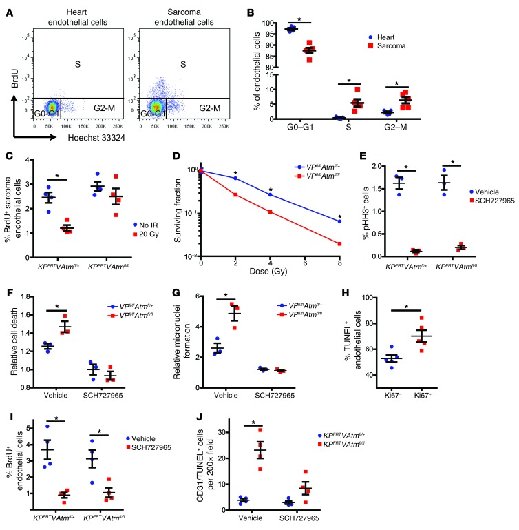
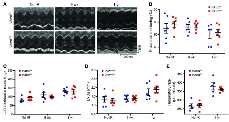
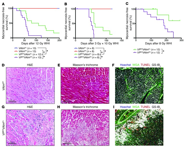
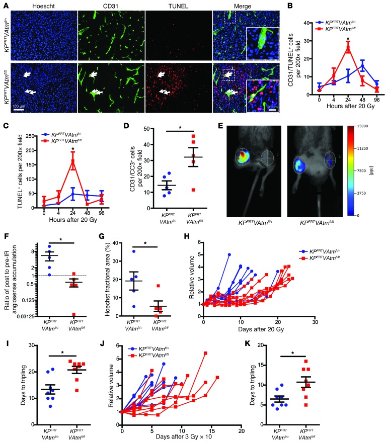
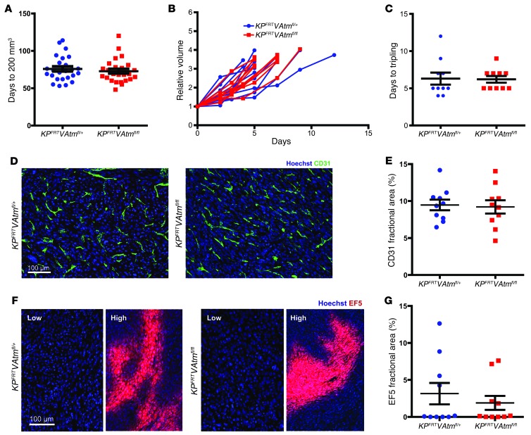
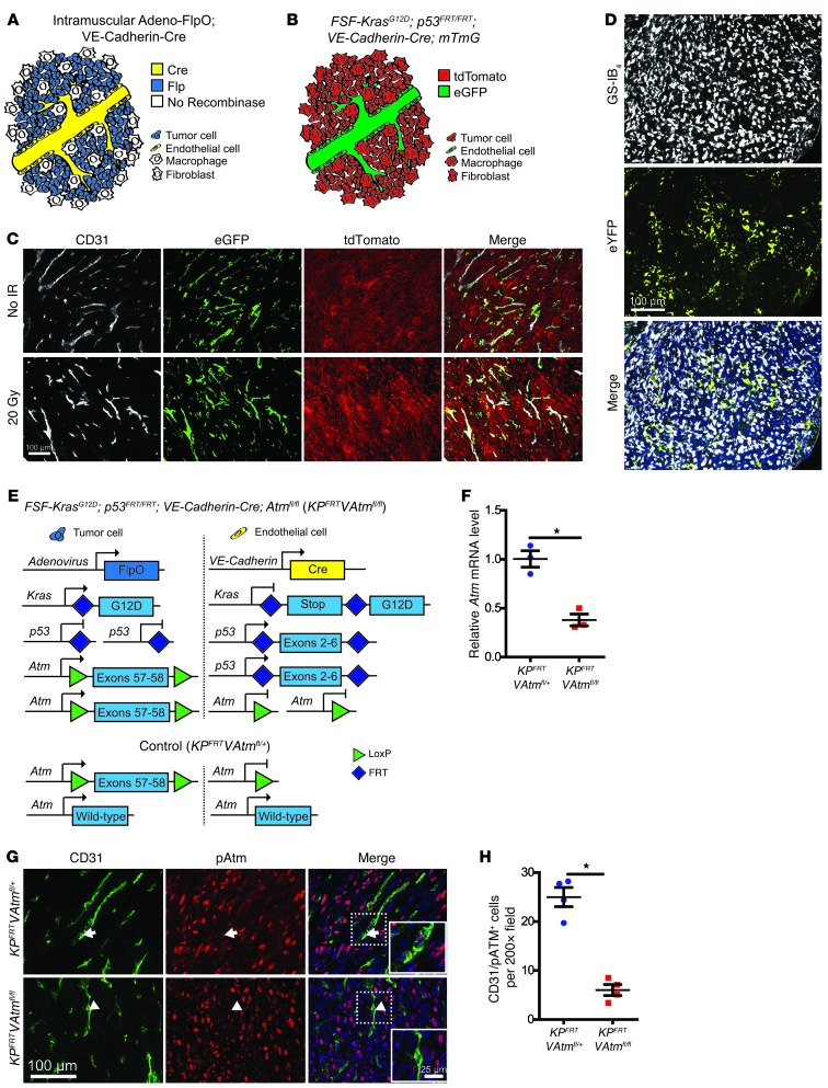
Cells isolated from patients with ataxia telangiectasia are exquisitely sensitive to ionizing radiation. Kinase inhibitors of ATM, the gene mutated in ataxia telangiectasia, can sensitize tumor cells to radiation therapy, but concern that inhibiting ATM in normal tissues will also increase normal tissue toxicity from radiation has limited their clinical application. Endothelial cell damage can contribute to the development of long-term side effects after radiation therapy, but the role of endothelial cell death in tumor response to radiation therapy remains controversial. Here, we developed dual recombinase technology using both FlpO and Cre recombinases to generate primary sarcomas in mice with endothelial cell-specific deletion of Atm to determine whether loss of Atm in endothelial cells sensitizes tumors and normal tissues to radiation. Although deletion of Atm in proliferating tumor endothelial cells enhanced the response of sarcomas to radiation, Atm deletion in quiescent endothelial cells of the heart did not sensitize mice to radiation-induced myocardial necrosis. Blocking cell cycle progression reversed the effect of Atm loss on tumor endothelial cell radiosensitivity. These results indicate that endothelial cells must progress through the cell cycle in order to be radiosensitized by Atm deletion.


Figures

Figure 6
Figure 6. Atm deletion sensitizes proliferating endothelial cells to radiation.
(A and B) Flow cytometry analysis (A) and quantification of cell cycle phase (B) in heart and sarcoma endothelial cells from KPFRT mice (n = 5). (C) Flow cytometry quantification of BrdU incorporation into tumor endothelial cells from KPFRTVAtmfl/+ and KPFRTVAtmfl/fl mice 1 hour after irradiation with 20 Gy or in unirradiated controls (n = 4 per group). (D) Clonogenic assay of primary cardiac endothelial cells from VPfl/flAtmfl/+ and VPfl/flAtmfl/fl mice (n = 3 independent experiments). (E) Flow cytometry quantification of phosphorylated histone H3 (pHH3) for primary cardiac endothelial cells from VPfl/flAtmfl/+ and VPfl/flAtmfl/fl mice treated with DMSO vehicle or 500 nM SCH727965 for 24 hours. (F and G) Cell death (F) and micronuclei formation (G), 24 hours after irradiation of primary cardiac endothelial cells from VPfl/flAtmfl/+ and VPfl/flAtmfl/fl mice treated with DMSO or 500 nM SCH727965 immediately before irradiation with 12 Gy (n = 3 independent experiments). Data are expressed relative to unirradiated cells of the same genotype and drug treatment. (H) Quantification of TUNEL staining in Ki67+ and Ki67 endothelial cells (CD31+) from tumors in KPFRTVAtmfl/fl mice 24 hours after irradiation with 20 Gy (n = 5). (I) Flow cytometry quantification of BrdU incorporation into sarcoma endothelial cells from KPFRTVAtmfl/+ and KPFRTVAtmfl/fl mice 24 hours after injection with vehicle or 40 mg/kg SCH727965 (n = 4 per group). (J) Quantification of CD31+TUNEL+ cells in sarcomas from KPFRTVAtmfl/+ and KPFRTVAtmfl/fl mice 24 hours after treatment with vehicle or SCH727965 immediately before irradiation with 20 Gy (n = 4 per group). All data are mean ± SEM. *P < 0.05.
Figure 5
Figure 5. Characterization of cardiac and respiratory function in VAtmfl/+ and VAtmfl/fl mice with and without whole-heart irradiation.
(A) Representative echocardiography recordings from VAtmfl/+ and VAtmfl/fl mice 6 weeks or 1 year after 12 Gy whole-heart irradiation and in unirradiated controls. (BD) Changes in fractional shortening (B), left ventricular mass (C), and left ventricular end-systolic dimension (LVDs) (D) in VAtmfl/+ and VAtmfl/fl mice 6 weeks and 1 year after 12 Gy whole-heart irradiation and in unirradiated controls (n = 6 mice per group). (E) Respiratory rate in VAtmfl/+ and VAtmfl/fl mice 1 year after 12 Gy whole-heart irradiation and in unirradiated controls (n = 5 per group). All data are mean ± SEM.
Figure 4
Figure 4. Deletion of Atm in p53 WT endothelial cells does not sensitize mice to radiation-induced myocardial necrosis.
(A) Kaplan-Meier plots of myocardial necrosis–free survival for VAtmfl/+, VAtmfl/fl, VPfl/flAtmfl/+, and VPfl/flAtmfl/fl mice after 12 Gy whole-heart irradiation. (B) Kaplan-Meier plots of myocardial necrosis–free survival for VAtmfl/+, VAtmfl/fl, VPfl/flAtmfl/+, and VPfl/flAtmfl/fl mice after whole-heart irradiation with 10 daily fractions of 3 Gy. 1 VPfl/flAtmfl/fl mouse died prior to finishing irradiation and was censored. (C) Kaplan-Meier plots of myocardial necrosis–free survival for VPfl/flAtmfl/+ and VPfl/flAtmfl/fl mice after 8 Gy whole-heart irradiation. Mice of both genotypes were censored due to development of thymic lymphomas prior to heart disease. (DI) Representative sections of the myocardium of (DF) a VAtmfl/fl mouse 469 days after whole-heart irradiation and of (GI) a VPfl/flAtmfl/+ mouse 56 days after whole-heart irradiation, subjected to staining with H&E (D and G) or Masson trichrome (E and H) or immunofluorescence for WGA, TUNEL, and GS-IB4 (F and I). *P < 0.05. Scale bars: 100 μm (DI).
Figure 3
Figure 3. Loss of Atm sensitizes tumor endothelial cells to ionizing radiation and increases the radiation response of primary sarcomas.
(A) Immunofluorescence for CD31 and TUNEL in sarcomas from KPFRTVAtmfl/+ and KPFRTVAtmfl/fl mice 24 hours after irradiation with 20 Gy. Examples of dead endothelial cells (white arrows) are shown at higher magnification in the insets. (B and C) Quantification of CD31+TUNEL+ cells (B) and total TUNEL+ cells (C) in sarcomas from KPFRTVAtmfl/+ and KPFRTVAtmfl/fl mice at various time points after irradiation with 20 Gy (n = 5 per group). (D) Quantification of CD31+CC3+ cells in sarcomas from KPFRTVAtmfl/+ and KPFRTVAtmfl/fl mice 24 hours after irradiation with 20 Gy (n = 5 per group). (E) FMT of the blood pool imaging agent AngioSense, injected 24 hours after irradiation of sarcomas from KPFRTVAtmfl/+ and KPFRTVAtmfl/fl mice with 20 Gy. (F) Quantification of the change in AngioSense accumulation after irradiation of sarcomas in KPFRTVAtmfl/+ and KPFRTVAtmfl/fl mice with 20 Gy (n = 5 per group). (G) Quantification of Hoechst 33342 perfusion in sarcomas from KPFRTVAtmfl/+ and KPFRTVAtmfl/fl mice 24 hours after irradiation with 20 Gy (n = 5 per group). (HK) Tumor growth curves and time to tripling for sarcomas in KPFRTVAtmfl/+ and KPFRTVAtmfl/fl mice after irradiation with 20 Gy (H and I) or 10 daily fractions of 3 Gy (J and K) (n = 8 per group). All data are mean ± SEM. *P < 0.05. Scale bars: 100 μm (A); 25 μm (A, insets).
Figure 2
Figure 2. Deletion of Atm in endothelial cells does not affect tumor growth or vascular development of primary soft tissue sarcomas.
(A) Time for sarcomas to reach 200 mm3 after intramuscular injection of adeno-FlpO in KPFRTVAtmfl/+ and KPFRTVAtmfl/fl mice (n = 25 per group). (B) Tumor growth curves and (C) time to tripling for unirradiated sarcomas in KPFRTVAtmfl/+ and KPFRTVAtmfl/fl mice (n = 10 per group). (D and E) Immunofluorescence (D) and quantification (E) of endothelial cell marker CD31 in sarcomas from KPFRTVAtmfl/+ and KPFRTVAtmfl/fl mice (n = 10 per group). (F and G) Immunofluorescence (F) and quantification (G) of hypoxia marker EF5 in sarcomas from KPFRTVAtmfl/+ and KPFRTVAtmfl/fl mice (n = 10 per group). Regions of low and high hypoxia are shown for both genotypes. All data are mean ± SEM. Scale bars: 100 μm (D and F).
Figure 1
Figure 1. Dual recombinase technology enables VE-Cadherin-Cre to delete Atm in primary sarcoma endothelial cells.
(A) Recombinase expression in KPFRT; VE-Cadherin-Cre mice injected with adeno-FlpO to generate sarcomas. (B) Reporter expression in KPFRT; VE-Cadherin-Cre; mTmG mice. All cells initially express tdTomato, and VE-Cadherin-Cre deletes tdTomato and turns on eGFP expression in endothelial cells (green). (C) Fluorescence images of CD31-stained soft tissue sarcomas initiated with adeno-FlpO in KPFRT; VE-Cadherin-Cre; mTmG mice in the absence of radiation (No IR) and 2 weeks after irradiation with 20 Gy. Images are representative of 3 mice per group. (D) Representative immunofluorescence images of a sarcoma in a KPloxP; LSL-eYFP mouse initiated with adeno-Cre and stained with GS-IB4. (E) Genetic strategy to activate Kras and delete p53 in tumor cells and delete Atm in endothelial cells. Control mice retained 1 WT allele of Atm in endothelial cells. (F) Expression of Atm mRNA in FACS-isolated tumor endothelial cells (CD45CD34+CD31+) from the indicated mice (n = 3 per group). (G and H) Immunofluorescence (G) and quantification (H) of CD31+pATM+ cells in sarcomas from KPFRTVAtmfl/+ and KPFRTVAtmfl/fl mice 4 hours after irradiation with 20 Gy (n = 4 per group). A pATM+ endothelial cell in the KPFRTVAtmfl/+ mouse (arrows) and a pATM endothelial cell in the KPFRTVAtmfl/fl mouse (arrowheads) are shown at higher magnification in the insets. Data are mean ± SEM. Scale bars: 100 μm (C, D, and G); 25 μm (G, insets) *P < 0.05.

Comment in

Similar articles

Cited by

References

    1. Delaney G, Jacob S, Featherstone C, Barton M. The role of radiotherapy in cancer treatment: estimating optimal utilization from a review of evidence-based clinical guidelines. Cancer. 2005;104(6):1129–1137. doi: 10.1002/cncr.21324. - DOI - PubMed
    1. Begg AC, Stewart FA, Vens C. Strategies to improve radiotherapy with targeted drugs. Nat Rev Cancer. 2011;11(4):239–253. doi: 10.1038/nrc3007. - DOI - PubMed
    1. Moding EJ, Kastan MB, Kirsch DG. Strategies for optimizing the response of cancer and normal tissues to radiation. Nat Rev Drug Discov. 2013;12(7):526–542. doi: 10.1038/nrd4003. - DOI - PMC - PubMed
    1. Shiloh Y. ATM and related protein kinases: safeguarding genome integrity. Nat Rev Cancer. 2003;3(3):155–168. doi: 10.1038/nrc1011. - DOI - PubMed
    1. Kastan MB, Lim DS. The many substrates and functions of ATM. Nat Rev Mol Cell Biol. 2000;1(3):179–186. doi: 10.1038/35043058. - DOI - PubMed

Publication types

MeSH terms

Substances