. 2014 Jul 18;9(7):e102077.
doi: 10.1371/journal.pone.0102077. eCollection 2014.

Hair cell regeneration after ATOH1 gene therapy in the cochlea of profoundly deaf adult guinea pigs

Affiliations

Hair cell regeneration after ATOH1 gene therapy in the cochlea of profoundly deaf adult guinea pigs

Patrick J Atkinson et al. PLoS One. .

Abstract

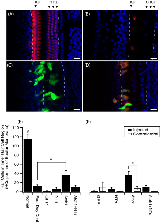
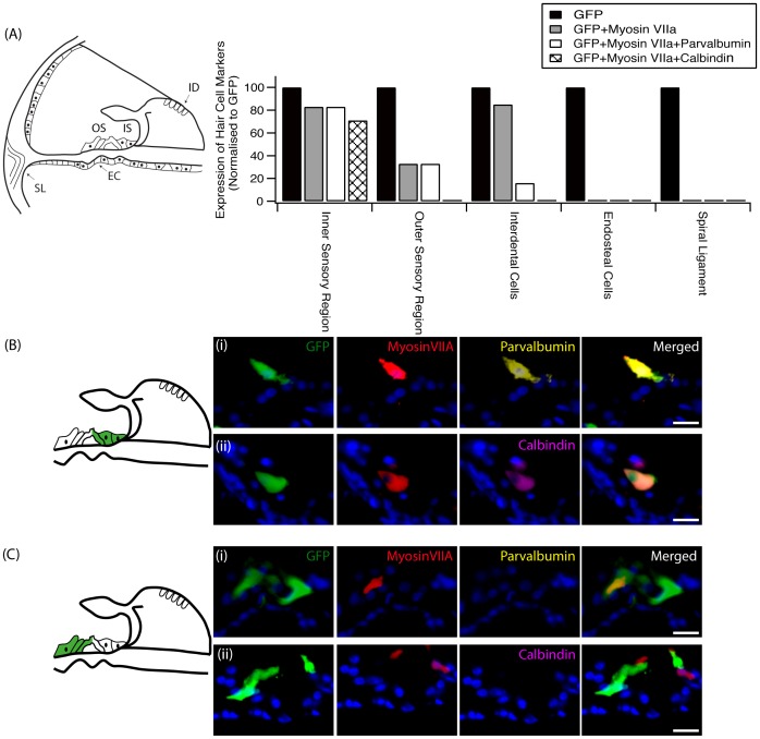
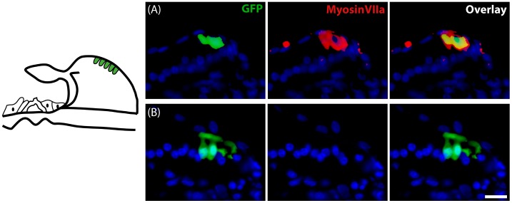
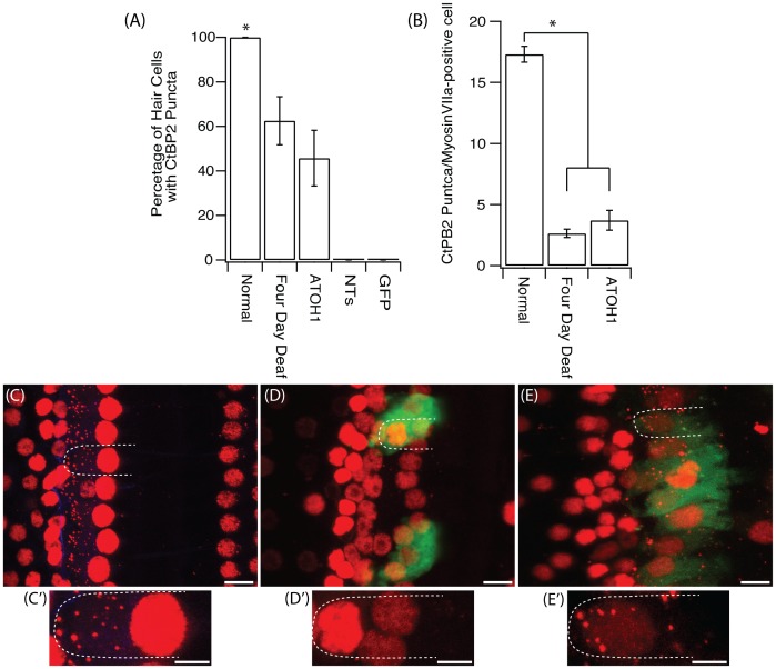
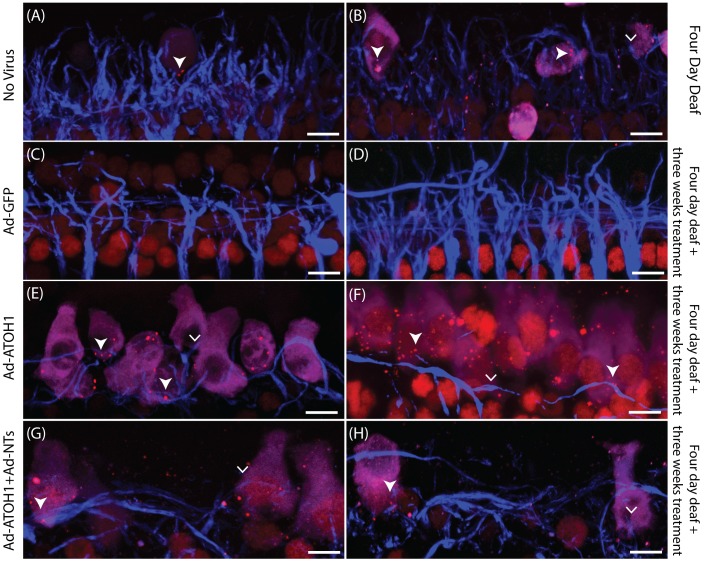
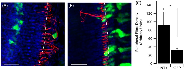
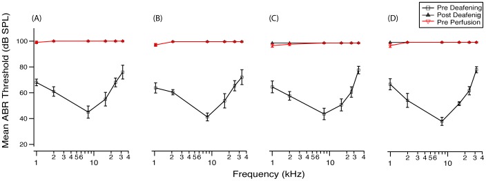
The degeneration of hair cells in the mammalian cochlea results in permanent sensorineural hearing loss. This study aimed to promote the regeneration of sensory hair cells in the mature cochlea and their reconnection with auditory neurons through the introduction of ATOH1, a transcription factor known to be necessary for hair cell development, and the introduction of neurotrophic factors. Adenoviral vectors containing ATOH1 alone, or with neurotrophin-3 and brain derived neurotrophic factor were injected into the lower basal scala media of guinea pig cochleae four days post ototoxic deafening. Guinea pigs treated with ATOH1 gene therapy, alone, had a significantly greater number of cells expressing hair cell markers compared to the contralateral non-treated cochlea when examined 3 weeks post-treatment. This increase, however, did not result in a commensurate improvement in hearing thresholds, nor was there an increase in synaptic ribbons, as measured by CtBP2 puncta after ATOH1 treatment alone, or when combined with neurotrophins. However, hair cell formation and synaptogenesis after co-treatment with ATOH1 and neurotrophic factors remain inconclusive as viral transduction was reduced due to the halving of viral titres when the samples were combined. Collectively, these data suggest that, whilst ATOH1 alone can drive non-sensory cells towards an immature sensory hair cell phenotype in the mature cochlea, this does not result in functional improvements after aminoglycoside-induced deafness.


Conflict of interest statement

Competing Interests: The authors have declared that no competing interests exist.

Figures

Figure 1
Figure 1. An increase in hair cells in the inner hair cell region after ATOH1 gene therapy.
Example photomicrographs of surface preparations of the basal turn of the cochlea from (A) normal, (B) four day deaf, (C) Ad-GFP treated, (D) Ad-ATOH1 treated animals (red  =  myosinVIIa, green  =  GFP, blue  =  DAPI, dashed lines mark the sensory region, scale bar  = 20 µm). (E) When quantified there was a significant loss of IHCs after four days of deafness compared to normal hearing animals. Three weeks post ATOH1-gene therapy there were significantly more myosinVIIa positive cells in the IHC region compared with the four day deaf group (*p<0.05, ANOVA). This number however remained below that observed in normal cochleae which had a greater number of IHCs compared to any other treatment group (#p<0.05, ANOVA). (F) A comparison of treated (injected) versus non-treated (contralateral) cochleae showed ATOH1-injected cochleae to have a significantly greater number of IHCs relative to the contralateral control cochleae (§p<0.05 paired t-test).
Figure 2
Figure 2. HC marker expression following ATOH1 treatment.
(A) The transduction of cells with ATOH1 resulted in cells expressing HC markers to varying degrees depending on their location (EC: endosteal cells, ID: interdental cells, IS: inner sensory region, OS: outer sensory region, SL: spiral ligament). Transduced cells (green) in the inner sensory region (B) had the highest percentage of cells that expressed the HC markers myosinVIIa (red), parvalbumin (yellow) and calbindin (magenta). A high proportion of myosinVIIa-positive transduced cells in this region co-labelled with (i) parvalbumin or (ii) calbindin. Fewer cells in the outer sensory region (C) expressed HC markers. ATOH1-transduced cells within the spiral ligament or endosteal cell region did not express any HC markers. Scale bar  = 20 µm.
Figure 3
Figure 3. Transduction of interdental cells following ATOH1 or GFP treatment.
Schematic at left illustrates the region of the interdental cells within the spiral limbus. The transduction of interdental cells (green) with ATOH1 (A) resulted in the expression of the HC marker myosinVIIa (red) in 85% of the cells. (B) MyosinVIIa expression was not detected following transduction of interdental cells with the GFP control vector. Scale bar  = 20 µm.
Figure 4
Figure 4. Fure 4. CtBP2 expression in IHCs.
(A) There was a significant decrease in the percentage of HCs that were positive for CtBP2 puncta after four days of deafness and after treatment with ATOH1 when compared to the normal hearing cochlea (*p<0.05, one way ANOVA). (B) There were also significantly less CtBP2 puncta per IHC after four days of deafness or ATOH1 treatment. Examples of CtBP2 staining (red) in the (C) normal, (D) GFP treated and (E) ATOH1 treated cochlea with their respective magnified image (C′-E′), green  =  GFP. Scale bar  = 10 µm (main image) and 5 µm (magnified image)
Figure 5
Figure 5. Surface preparations illustrating peripheral fibres in close proximity to HCs with ribbon synapses.
Examples of peripheral fibres (blue) close to myosinVIIa-positive cells (magenta) with CtBP2 puncta (red; filled arrowhead) or without CtBP2 puncta (arrowhead) in the four day deafened (A&B), GFP treated (C&D), ATOH1 treated (E&F) or ATOH1+NTs (G&H) treated cochleae. Scale bar  = 10 µm.
Figure 6
Figure 6. Resprouting peripheral fibres.
Cochlear surface preparations were stained with anti-NF (red) and DAPI (blue). (A) Peripheral fibre responses to NT-expressing cells in the OC post Ad-NTs treatment. (B) Peripheral fibre response to GFP only expressing cells in the OC post Ad-GFP treatment. The perforated lines indicate the approximate medial edge of the inner pillar cells of the OC. Scale bar  = 50 µm. (C) Significantly greater fibre density was observed in close proximity (≤10 µm) to NT-secreting cells compared to GFP-only transduced cells (p<0.05, t-test).
Figure 7
Figure 7. Mean ABR thresholds pre-deafening, post-deafening and post-treatment.
There was a profound hearing loss across all frequencies measured (1, 2, 8, 16, 24, 32 kHz) four days post aminoglycoside deafening for all animals. Three weeks after treatment with either (A) Ad-GFP, (B) Ad-NTs, (C) Ad-ATOH1 or (D) Ad-ATOH1+Ad-NTs there was no change in hearing thresholds compared to those measured post-deafening (p>0.05, repeated measured ANOVA).

References

    1. Feghali JG, Lefebvre PP, Staecker H, Kopke R, Frenz DA, et al. (1998) Mammalian auditory hair cell regeneration/repair and protection: a review and future directions. Ear Nose Throat J. 77: 276, 280, 282–275. - PubMed
    1. Fu QJ, Nogaki G (2005) Noise susceptibility of cochlear implant users: the role of spectral resolution and smearing. J Assoc Res Otolaryngol 6: 19–27. - PMC - PubMed
    1. Kong YY, Cruz R, Jones JA, Zeng FG (2004) Music perception with temporal cues in acoustic and electric hearing. Ear Hear 25: 173–185. - PubMed
    1. Kelly MC, Chen P (2009) Development of form and function in the mammalian cochlea. Curr Opin Neurobiol 19: 395–401. - PMC - PubMed
    1. Bermingham NA, Hassan BA, Price SD, Vollrath MA, Ben-Arie N, et al. (1999) Math1: an essential gene for the generation of inner ear hair cells. Science 284: 1837–1841. - PubMed

Publication types

Substances