Case Reports
. 2014 Aug 19;83(8):743-51.
doi: 10.1212/WNL.0000000000000716. Epub 2014 Jul 18.

Mitochondrial EFTs defects in juvenile-onset Leigh disease, ataxia, neuropathy, and optic atrophy

Affiliations
Case Reports

Mitochondrial EFTs defects in juvenile-onset Leigh disease, ataxia, neuropathy, and optic atrophy

Sofia Ahola et al. Neurology. .

Abstract

Objective: We report novel defects of mitochondrial translation elongation factor Ts (EFTs), with high carrier frequency in Finland and expand the manifestations of this disease group from infantile cardiomyopathy to juvenile neuropathy/encephalopathy disorders.

Methods: DNA analysis, whole-exome analysis, protein biochemistry, and protein modeling.

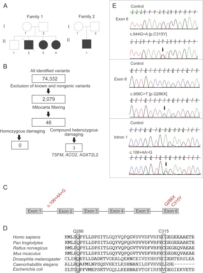
Results: We used whole-exome sequencing to find the genetic cause of infantile-onset mitochondrial cardiomyopathy, progressing to juvenile-onset Leigh syndrome, neuropathy, and optic atrophy in 2 siblings. We found novel compound heterozygous mutations, c.944G>A [p.C315Y] and c.856C>T [p.Q286X], in the TSFM gene encoding mitochondrial EFTs. The same p.Q286X variant was found as compound heterozygous with a splice site change in a patient from a second family, with juvenile-onset optic atrophy, peripheral neuropathy, and ataxia. Our molecular modeling predicted the coding-region mutations to cause protein instability, which was experimentally confirmed in cultured patient cells, with mitochondrial translation defect and lacking EFTs. Only a single TSFM mutation has been previously described in different populations, leading to an infantile fatal multisystem disorder with cardiomyopathy. Sequence data from 35,000 Finnish population controls indicated that the heterozygous carrier frequency of p.Q286X change was exceptionally high in Finland, 1:80, but no homozygotes were found in the population, in our mitochondrial disease patient collection, or in an intrauterine fetal death material, suggesting early developmental lethality of the homozygotes.

Conclusions: We show that in addition to early-onset cardiomyopathy, TSFM mutations should be considered in childhood and juvenile encephalopathies with optic and/or peripheral neuropathy, ataxia, or Leigh disease.


Figures

Figure 1
Figure 1. Exome sequencing revealed 3 novel mutations in the TSFM gene in 2 families
(A) The pedigrees. Black symbols indicate affected individuals; strike through indicates deceased subject. (B) Exome data analysis and filtering steps of the single nucleotide variants of patient 1 (P1, family 1, II-3). TSFM, AGXT2L2 (PHYKPL, 5-phosphohydroxy-l-lysine phospho-lyase) and ACO2 (aconitase 2) indicate the genes in which potentially pathogenic variants were identified. TSFM gave best variant calling quality and was thus selected for further analysis. (C) TSFM gene structure with all 3 identified mutations marked in red. (D) Conservation of mutant elongation factor Ts amino acids Q286 and C315 in species. (E) Chromatograms of the 3 identified mutations (arrows). All mutations were confirmed to be heterozygous.
Figure 2
Figure 2. TSFM mutations lead to protein degradation and mild reduction in translation activity
(A) Relative messenger RNA expressions of EFTs in cultured cells of P1, P2, and P3. (B) Structural modeling of EFTu-EFTs complex showing the 2 amino acid changes p.Q286X (deleted sequence in yellow) and p.C315Y. Color coding: gray = EFTu; magenta = residues forming the GDP binding site in EFTu; pink = EFTs amino terminal domain (residues 1–99); green = the core domain (residues 100–210); blue = subdomain C (residues 211–318); and red = EFTs interaction site with EFTu and nucleotide exchange. p.C315 fits tightly into an uncharged pocket predicted to be formed by the residues from the opposite side of the β-sandwich. Substitution of cysteine 315 for tyrosine introduces a bulky side chain into the pocket designed for cysteine, resulting in van der Waals repulsion between the halves of the β-sandwich oriented toward the EFTu contact surface. This change in the local architecture affects stability of EFTs. Structural rearrangements caused by the mutation will affect the position of p.E104 locating in a distance of 3.8 Å from p.D126, which forms part of the α-turn in the subdomain N (marked in red) critical for interaction with EFTu and nucleotide exchange. (C) Immunoblot analysis showed almost complete loss of EFTs protein in P1 and P2 fibroblasts and substantial reduction in P3 myoblasts. Mild EFTu reduction was also seen in P1 and P2 but not in P3 samples. (D) Mitochondrial translation was partially reduced in P2 fibroblasts, but suggestively P1. An evident reduction of translation was seen in P3 myoblasts. R312W = patient fibroblast cell line with previously published EFTs mutation as a positive control for translation defect. ATP = ATP synthase; CO = cytochrome c oxidase; cytb = cytochrome b; EFTs = elongation factor Ts; EFTu = elongation factor Tu; GDP = guanosine diphosphate; ND = NADH dehydrogenase; SDH = succinate dehydrogenase.

References

    1. Ylikallio E, Suomalainen A. Mechanisms of mitochondrial diseases. Ann Med 2012;44:41–59 - PubMed
    1. Antonicka H, Sasarman F, Kennaway NG, Shoubridge EA. The molecular basis for tissue specificity of the oxidative phosphorylation deficiencies in patients with mutations in the mitochondrial translation factor EFG1. Hum Mol Genet 2006;15:1835–1846 - PubMed
    1. Calvo SE, Compton AG, Hershman SG, et al. Molecular diagnosis of infantile mitochondrial disease with targeted next-generation sequencing. Sci Transl Med 2012;4:118ra110 - PMC - PubMed
    1. Coenen MJ, Antonicka H, Ugalde C, et al. Mutant mitochondrial elongation factor G1 and combined oxidative phosphorylation deficiency. N Engl J Med 2004;351:2080–2086 - PubMed
    1. Smeitink JA, Elpeleg O, Antonicka H, et al. Distinct clinical phenotypes associated with a mutation in the mitochondrial translation elongation factor EFTs. Am J Hum Genet 2006;79:869–877 - PMC - PubMed

Publication types

LinkOut - more resources