. 2014 Aug;141(16):3143-52.
doi: 10.1242/dev.106989. Epub 2014 Jul 18.

Tcf3 expression marks both stem and progenitor cells in multiple epithelia

Affiliations

Tcf3 expression marks both stem and progenitor cells in multiple epithelia

Jeffrey M Howard et al. Development. 2014 Aug.

Abstract

The Lef/Tcf-family transcription factor Tcf3 has important roles in development, stem cell function and malignancy. Previous gain- and loss-of-function studies have suggested that Tcf3 is a mediator of self-renewal and an undifferentiated state in stem and progenitor cells in skin, but little is known of its role in other postnatal tissues. Here, we explore the distribution and behavior of Tcf3-expressing cells in several adult tissues using a novel Tcf3-CreER knock-in mouse model. By lineage tracing in dorsal skin, we verify that Tcf3-expressing cells in the hair follicle bulge are self-renewing stem cells with multilineage potential. We then demonstrate, for the first time, the presence of Tcf3-expressing cells in the basal layer of several other stratified epithelia, including the paw skin, tongue and esophagus. By lineage tracing, we demonstrate that the Tcf3-expressing population in these tissues includes persistent stem cells, transient progenitors and cells undergoing active differentiation. Our observations here suggest that the role of Tcf3 in cell-fate decision is more complex than previously appreciated and is highly dependent on cellular context.

Keywords: Adult stem cells; Lineage tracing; Mouse; Tcf transcription factors; Transcription factor 7-like 1 protein.


Figures

Fig. 1.
Fig. 1.
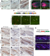
The Tcf3-2A-eGFP-2A-CreERT2 mouse is a faithful, tightly regulated reporter of Tcf3 expression in vivo. (A-C) Tcf3 immunohistochemistry in wild-type mouse dorsal skin. (A) In the telogen (resting) phase, Tcf3 is expressed in the hair follicle bulge (Bu) and dermal papilla (DP), and is absent from the sebaceous gland (SG) and interfollicular epidermis (IFE). (B,C) During anagen, Tcf3 expression is upregulated in the bulge and is expanded into the outer root sheath (ORS) of the growing hair, but is absent from the transit-amplifying cells of the matrix (Mx). Me, endogenous melanin pigmentation. (D,E) Immunofluorescent co-staining for Tcf3 and either Ki67 (B) or Lef1 (C) in wild-type anagen-phase skin. There is a clear division between Tcf3-expressing, non-proliferating ORS cells and Lef1-expressing, highly proliferative matrix cells. (F) Schematic of the Tcf3-2A-eGFP-2A-CreERT2 (‘Tcf3 GC’) knock-in allele. (G-I) Immunofluorescent co-staining for Tcf3 and GFP in neonatal knock-in and wild-type back skin. As expected, GFP colocalizes with Tcf3 in the developing hair follicles (arrowheads). (J-R) Immunohistochemical staining for GFP in Tcf3+/+ (J-L), GC/+ (M-O) and GC/GC (P-R) adult mouse skin in telogen and anagen phases of the hair cycle. GFP expression is observed in the hair follicle bulge (Bu), dermal papilla (DP) and outer root sheath (ORS), mirroring endogenous Tcf3 expression (compare A-C). SG, sebaceous gland (with nonspecific staining); Me, endogenous melanin. (S) Schematic of the ROSA26-mTmG Cre reporter allele used for lineage tracing. (T,U) Three days after treatment of Tcf3GC/+; ROSA26mTmG/+ mice with tamoxifen, individual Tcf3-lineage (Tcf3 lin) mGFP(+) cells are seen in the hair follicle bulge (arrowheads), as well as in various cell types within the dermis including blood vessels (T). No leaky Cre activity is observed in vehicle-treated controls (U). β4, β4 integrin. Scale bars: 100 µm in T,U; 50 µm in A-E,G-R.
Fig. 2.
Fig. 2.
Tcf3 expression marks hair follicle stem cells in murine dorsal skin. (A-K) Lineage traces in Tcf3-expressing hair follicle bulge cells. Tcf3GC/+; ROSA26mTmG/+ mice were treated with tamoxifen at P21 and P22 and sacrificed 3 days to 6 months after induction. (A,B) Individual Tcf3-lineage (Tcf3 lin) cells in the HF bulge (Bu) (A, arrowhead), hair germ (HG) and dermal papilla (DP) (B, arrowhead) 3 days after induction. Asterisk indicates hair shaft autofluorescence. (C-H) Two weeks after induction, Tcf3-lineage cells (arrowheads) are seen in the keratin 5 (K5)-positive outer root sheath (ORS) (C), Lef1-positive transit-amplifying matrix cells (Mx) (D) and all differentiated lineages of the hair shaft. Hair shaft lineage markers: companion layer (Cp) and keratin 6 (K6) (E); cortex (Cx) and hair cortex cytokeratin (AE13) (F); inner root sheath (IRS) and medulla (Me), trichohyalin (AE15) (G,H). (I-K) Six months after treatment, persistent clones of single and multiple cells are detected in the bulge (Bu) of both telogen- (I) and anagen-phase (J) hair follicles (arrowheads). In anagen skin, mGFP(+) clones expand and incorporate into growing hair shafts via the transit-amplifying hair matrix (Mx) (K). (L-O) Lineage traces in Tcf3-expressing cells during anagen. Tcf3GC/+; ROSA26mTmG/+ mice were treated with tamoxifen during full anagen (∼P30) and sacrificed 3 days or 4 weeks after induction. (L,M) Three days after induction, numerous Tcf3-lineage clones are seen in the outer root sheath of nearly all follicles (L); a minority of follicles also show labeling in the bulge (Bu) (M). (N) Four weeks after induction, when mice have re-entered telogen, most bulges contain mGFP(+) cells. (O) Quantification of the proportion of HFs containing mGFP(+) cells in the bulge region 3 days and 4 weeks after lineage-tracing induction during anagen. The analysis was performed in two ways: first, counting follicles with labeled cells in any portion of the bulge (dark circles/squares); second, counting only follicles containing cells in the outer layer of the bulge (open circles/squares). Only a minority of bulge regions (16.5±7.2% for total bulge, 15.9±8.3% for outer bulge) contain mGFP(+) cells at the initial timepoint. The proportion rises to 70.2±16.6% (total bulges) or 44.0±10.3% (outer bulges) after follicles have returned to telogen (all percentages mean±s.d.; P=0.0095 for both comparisons, Mann–Whitney U-test). Data points represent percentage of HF bulge regions containing mGFP(+) cells in individual mice; bars indicate mean. β4, β4 integrin. Scale bars: 100 µm in L; 50 µm in A-K,M,N.
Fig. 3.
Fig. 3.
Tcf3-expressing hair follicle bulge cells contribute to wound repair of the interfollicular epithelium, but most fail to persist long term. Tcf3 lineage-tracing experiments in wounded epithelia. Tcf3GC/GC; ROSA26mTmG/+ mice were treated with tamoxifen at P21, P23 and P25, wounded at P30, and sacrificed 1-4 weeks after wounding. (A-E) Random cryosections of wound epithelium 1-4 weeks after wounding. (A) One week after wounding, Tcf3-lineage (Tcf3 lin) cells are observed migrating out of wound-adjacent hair follicles and incorporating into the wound epithelium (WE). (B) Tcf3-lineage cells (arrowheads) in the wound epithelium are primarily seen in the suprabasal layers. Numerous Tcf3-lineage cells are also seen in the wound dermis (Der). (C) By 3-4 weeks post wounding, the majority of Tcf3-lineage cells are lost from the wound epithelium but persist in the dermis. (D,E) A subset of Tcf3-lineage cells (arrowheads) does persist in the wound epithelium, frequently (E) associated with wound-adjacent hair follicles (HF). (F) Quantification of Tcf3-lineage clones in wound epithelium 1 and 4 weeks after wounding. Dots indicate number of mGFP(+) cells per 10 mm of wound epithelium in random cryosections at each timepoint; bars indicate mean. There is a trend towards cell loss over time, with only a minority (22.8±10.4%, mean±s.d.) of labeled cells persisting for at least 4 weeks (P=0.093, Mann–Whitney U-test). (G,H) Images of intact wound epithelium in Tcf3 lineage-tracing mice 2 (G) and 4 (H) weeks after wounding. Rare clones of mGFP(+) cells (arrowheads) are grossly visible in the wound epithelium, many of which are absent at the later timepoint. Tcf3 lin, Tcf3 lineage; β4, β4 integrin. Scale bars: 100 µm in A-C; 50 µm in D,E; 1 mm in G,H.
Fig. 4.
Fig. 4.
Tcf3 expression marks stem and progenitor cells in unhaired paw skin. (A-C) Immunofluorescent co-staining for Tcf3 and basal marker keratin 5 (K5) in wild-type plantar paw skin. (A,B) Tcf3 expression is seen throughout the basal layer of the stratified paw skin and, at lower levels, in occasional suprabasal cells (B, arrowhead). (C) Tcf3 is most highly expressed in the thick skin overlying the digital pads (DiP) (arrowheads). (D-L) Lineage tracing in paw skins of Tcf3GC/+; ROSA26mTmG/+ mice. Mice were treated with tamoxifen at P21 and P22 and sacrificed 3 days to 6 months after induction. (D) Three days post induction, single Tcf3-lineage (Tcf3 lin) cells are labeled in the basal layer. β4, β4 integrin. (E-H) Over time, mGFP(+) clones gave rise to columns of differentiated cells occupying the full thickness of the epithelium; some clones persisted for at least 6 months in vivo (H). (I-L) Epithelial layers and markers: basal layer, keratin 5 (K5) (I); spinous layer, keratin 1 (K1) (J) and filaggrin (Fil) (K); granular layer, loricrin (Lor) (L). (M) Extensive labeling was observed in hair follicles in adjacent haired paw skin. As in the dorsal skin, these were confined to the HF and never extended into the IFE. Scale bars: 20 µm in B; 100 µm in C; 50 µm in A,D-M.
Fig. 5.
Fig. 5.
Tcf3 expression marks stem and progenitor cells in esophageal mucosa. (A,B) Immunofluorescent co-staining for Tcf3 and basal marker keratin 5 (K5) in wild-type esophageal mucosa. Tcf3 is expressed throughout the basal layer and in some suprabasal cells (B, arrowhead). (C-J) Lineage tracing in esophageal mucosa of Tcf3GC/+; ROSA26mTmG/+ mice. Mice were treated with tamoxifen at P21/22 and sacrificed 3 days to 6 months after induction. (C) Three days post induction, small clones of 1-3 Tcf3-lineage (Tcf3 lin) cells were seen, primarily in the epithelial basal layer (arrowhead). β4, β4 integrin. (D-J) Over time, mGFP(+) clones formed columns of differentiated cells occupying the full thickness of the epithelium; some clones persisted for at least 6 months in vivo (H). Epithelial markers: suprabasal cells, keratin 13 (K13) (I); granular layer (Lor) (J). Scale bars: 20 µm in B; 50 µm in A,C-J.
Fig. 6.
Fig. 6.
Tcf3 expression marks stem and progenitor cells in lingual mucosa. (A,B) Immunofluorescent co-staining for Tcf3 and basal marker keratin 5 (K5) in wild-type dorsal tongue. Tcf3 expression is seen throughout the basal layer and at lower levels and in some suprabasal cells (B, arrowheads). (C-K) Lineage tracing in lingual epithelium of Tcf3GC/+; ROSA26mTmG/+ mice. Mice were treated with tamoxifen at P21 and P22 and sacrificed 3 days to 6 months after induction. (C) Three days after induction, clones of 2-5 mGFP(+) cells were seen in the epithelial basal layer (arrowhead). (D-K) Labeled clones expanded over time and incorporated into both filliform papilla (FP) and interpapillary pit (IPP) lineages. Cell type markers: interpapillary epithelium, keratin 13 (K13) (H); granular layer, loricrin (Lor) (I); filliform papillae, filaggrin (Fil) (J) and trichohyalin (AE15) (K). (L) The overwhelming majority of Tcf3-lineage clones were lost from the lingual epithelium over time, most within the first few weeks after induction. Clone frequency at 6 months post induction was only 10.0±5.1% (mean±s.d.) of the initial timepoint (P<0.001, Dunn's multiple comparison test). Dots indicate number of mGFP(+) clones per sagittal section of dorsal tongue in individual mice; bars indicate mean. (M-O) The dorsal surface of the tongue is covered by a dense layer of hook-shaped filliform papillae (FPs) in staggered rows. FPs are separated by interpapillary pits (IPPs) containing a distinct interpapillary epithelium (IPE). Long-term persistent clones occupied the entirety of a single interpapillary pit and incorporated into regions of the three or four neighboring filliform papillae, similar to previously described Bmi1-lineage clones (Tanaka et al., 2013). (M) Whole-mount image of intact dorsal epithelium from a P21 to 6 months lineage tracing tongue. Individual FPs are visible as triangular structures expressing the constitutive ROSA26-mT label. An individual mGFP(+) clone is seen, occupying a single IPP and contributing to each of the three neighboring FPs (arrowheads). (N,O) Sections cut through different planes of mGFP(+) clones in P21 to 6 months lineage-tracing tongue specimen, illustrating contribution of labeled cells to the anterior and posterior (N) or lateral (O) compartments of neighboring FPs. Insets represent a top-down schematic view of the dorsal tongue, showing the location of the section (red dashed line) within the labeled clone (green triangle) and relative to the neighboring FPs (conical structures). β4, β4 integrin. Scale bars: 20 µm in B; 200 µm in M; 50 µm in A,C-K,N,O.

References

    1. Ben-Porath I., Thomson M. W., Carey V. J., Ge R., Bell G. W., Regev A., Weinberg R. A. (2008). An embryonic stem cell-like gene expression signature in poorly differentiated aggressive human tumors. Nat. Genet. 40, 499-507 10.1038/ng.127 - DOI - PMC - PubMed
    1. Brownell I., Guevara E., Bai C. B., Loomis C. A., Joyner A. L. (2011). Nerve-derived sonic hedgehog defines a niche for hair follicle stem cells capable of becoming epidermal stem cells. Cell Stem Cell 8, 552-565 10.1016/j.stem.2011.02.021 - DOI - PMC - PubMed
    1. Cole M. F., Johnstone S. E., Newman J. J., Kagey M. H., Young R. A. (2008). Tcf3 is an integral component of the core regulatory circuitry of embryonic stem cells. Genes Dev. 22, 746-755 10.1101/gad.1642408 - DOI - PMC - PubMed
    1. Cotsarelis G., Sun T.-T., Lavker R. M. (1990). Label-retaining cells reside in the bulge area of pilosebaceous unit: implications for follicular stem cells, hair cycle, and skin carcinogenesis. Cell 61, 1329-1337 10.1016/0092-8674(90)90696-C - DOI - PubMed
    1. DasGupta R., Fuchs E. (1999). Multiple roles for activated LEF/TCF transcription complexes during hair follicle development and differentiation. Development 126, 4557-4568. - PubMed

Publication types

Substances