. 2014 Sep 15;307(6):H858-68.
doi: 10.1152/ajpheart.00308.2014. Epub 2014 Jul 18.

Systemic influences contribute to prolonged microvascular rarefaction after brain irradiation: a role for endothelial progenitor cells

Affiliations

Systemic influences contribute to prolonged microvascular rarefaction after brain irradiation: a role for endothelial progenitor cells

Nicole M Ashpole et al. Am J Physiol Heart Circ Physiol. .

Abstract

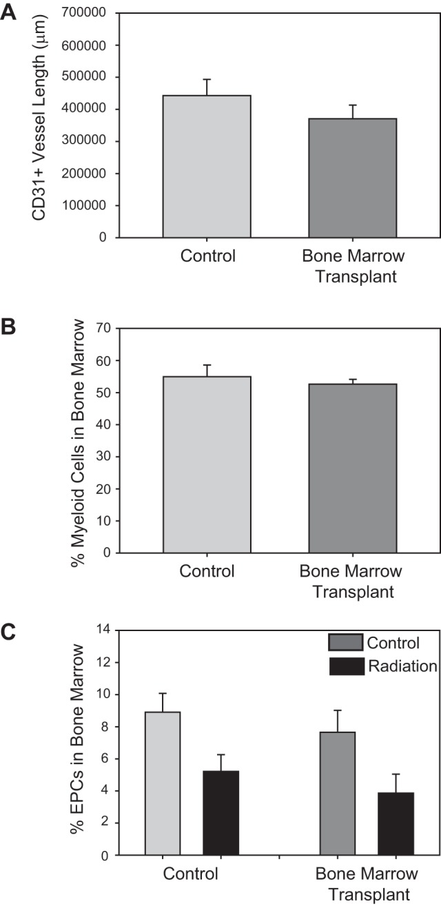
Whole brain radiation therapy (WBRT) induces profound cerebral microvascular rarefaction throughout the hippocampus. Despite the vascular loss and localized cerebral hypoxia, angiogenesis fails to occur, which subsequently induces long-term deficits in learning and memory. The mechanisms underlying the absence of vessel recovery after WBRT are unknown. We tested the hypotheses that vascular recovery fails to occur under control conditions as a result of loss of angiogenic drive in the circulation, chronic tissue inflammation, and/or impaired endothelial cell production/recruitment. We also tested whether systemic hypoxia, which is known to promote vascular recovery, reverses these chronic changes in inflammation and endothelial cell production/recruitment. Ten-week-old C57BL/6 mice were subjected to a clinical series of fractionated WBRT: 4.5-Gy fractions 2 times/wk for 4 wk. Plasma from radiated mice increased in vitro endothelial cell proliferation and adhesion compared with plasma from control mice, indicating that WBRT did not suppress the proangiogenic drive. Analysis of cytokine levels within the hippocampus revealed that IL-10 and IL-12(p40) were significantly increased 1 mo after WBRT; however, systemic hypoxia did not reduce these inflammatory markers. Enumeration of endothelial progenitor cells (EPCs) in the bone marrow and circulation indicated that WBRT reduced EPC production, which was restored with systemic hypoxia. Furthermore, using a bone marrow transplantation model, we determined that bone marrow-derived endothelial-like cells home to the hippocampus after systemic hypoxia. Thus, the loss of production and homing of EPCs have an important role in the prolonged vascular rarefaction after WBRT.

Keywords: angiogenesis; cytokines; endothelial progenitor cells; hypoxia; radiation therapy.


Figures

Fig. 1.
Fig. 1.
Experimental timeline. A and B: timelines for experiments using naïve mice (A) or mice with a bone marrow transplant (BMT; B). WBRT, whole brain radiation therapy; EPCs, endothelial progenitor cells.
Fig. 2.
Fig. 2.
WBRT-induced increases of the proangiogenic drive in the circulation. A: representative image of brain-derived microvascular endothelial cells (BDMECs) in culture 24 h after the addition of plasma from control (left) or radiated (right) mice. B: tube density within the BDMECs shown in A. C: cell adhesion after treatment of BDMECs with plasma from control or radiated mice. D: Time to reach 50% adhesion (T50 of cell adhesion) after plasma treatment. E: change in the proliferation of BDMECs after treatment with plasma from control or radiated mice, as measured using the Guava CellGrowth assay. F: fold changes in gene expression within BDMECs 24 h after plasma application. ANGPT, angiopoietin; VEGFR, VEGF receptor; SDF1, stromal cell-derived factor 1; MMP9, matrix metalloproteinase 9; THBS, thrombospondin. All results are means ± SE. *Significant difference compared with control (P < 0.05 by t-test).
Fig. 3.
Fig. 3.
Assessment of tissue inflammation in the hippocampus after WBRT and/or hypoxia. A and B: levels of IL-10 (A) and IL-12(p40) (B) after WBRT and/or systemic hypoxic treatment. All results are means ± SE. *Significant difference compared with control/normoxia (P < 0.05 by two-way ANOVA with a post hoc Bonferroni test).
Fig. 4.
Fig. 4.
Changes in EPCs within the bone marrow and blood after WBRT and hypoxia relative to control animals. A and B: bone marrow-derived EPCs (A) and EPCs within the blood (B) immediately after 4 wk of fractionated WBRT. C: fold changes in bone marrow-derived EPCs 2 mo after fractionated WBRT, with 2 wk of normoxia or hypoxia. D: EPCs in the blood 2 mo after WBRT, with 2 wk of normoxia or hypoxia. *Significant difference compared with control (normoxia); #significant difference between radiated normoxia and hypoxia (P < 0.05 by t-test).
Fig. 5.
Fig. 5.
Hypoxia induces a time-dependent mobilization of EPCs from the bone marrow. Fold change in EPCs within the circulation of control animals (left) and radiated animals (right) at various time points after the initiation of systemic hypoxia. Data are means ± SE. *Significant difference compared with normoxia (P < 0.05 by one-way ANOVA).
Fig. 6.
Fig. 6.
Basal effects of BMT. A: CD31+ vessel length in the hippocampus of control or BMT animals. B: percentages of myeloid cells in the bone marrow, as determined by flow cytometry. C: percentages of EPCs in the bone marrow of control or BMT mice 2 mo after WBRT or under control conditions. Data are means ± SE.
Fig. 7.
Fig. 7.
Bone marrow-derived EPCs home to the vasculature in the brain. A and B: representative images from the CA1 region of the hippocampus of radiated mice subjected to 2 wk of normoxia (A) or hypoxia (B). Red indicates CD31 staining, green indicates green fluorescent protein (GFP)+ bone marrow-derived cells (BMDCs), and blue indicates 4′,6-diamidino-2-phenylindole (DAPI) staining. C and D: Representative images from the CA1 region of the hippocampus when the head of BMT animals was shielded (C) or not shielded (D) during bone marrow treatment. Red indicates ionized Ca2+-binding adapter molecule 1 (Iba1) staining, green indicates GFP+ BMDCs, and blue indicates DAPI staining. E: GFP+ vessel length in the hippocampus of BMT animals 2 mo after control/WBRT and subsequent normoxia/hypoxia. Data are means ± SE. F: CD31+ vessel length in the hippocampus of control or BMT animals 2 mo after control/WBRT and subsequent normoxia/hypoxia. Data are means ± SE. *Significant difference between control and radiation; #significant difference between normoxia and hypoxia (P < 0.05).

References

    1. Ahn G, Brown J. Role of endothelial progenitors and other bone marrow-derived cells in the development of the tumor vasculature. Angiogenesis 12: 159–164, 2009 - PMC - PubMed
    1. Akiyama K, Tanaka R, Sato M, Takeda N. Cognitive dysfunction and histological findings in adult rats one year after whole brain irradiation. Neurol Med Chir (Tokyo) 41: 590–598, 2001 - PubMed
    1. Angiolillo AL, Sgadari C, Tosato G. A role for the interferon-inducible protein 10 in inhibition of angiogenesis by interleukin-12. Ann NY Acad Sci 795: 158–167, 1996 - PubMed
    1. Au P, Daheron L, Duda D, Cohen K, Tyrrell J, Lanning R, Fukumura D, Scadden D, Jain R. Differential in vivo potential of endothelial progenitor cells from human umbilical cord blood and adult peripheral blood to form functional long-lasting vessels. Blood 111: 1302–1305, 2008 - PMC - PubMed
    1. Caballero S, Sengupta N, Afzal A, Chang K, Li Calzi S, Guberski D, Kern T, Grant M. Ischemic vascular damage can be repaired by healthy, but not diabetic, endothelial progenitor cells. Diabetes 56: 960–967, 2007 - PMC - PubMed

Publication types

MeSH terms

LinkOut - more resources