. 2014 Sep 1;74(17):4741-51.
doi: 10.1158/0008-5472.CAN-14-1547. Epub 2014 Jul 18.

NACK is an integral component of the Notch transcriptional activation complex and is critical for development and tumorigenesis

Affiliations

NACK is an integral component of the Notch transcriptional activation complex and is critical for development and tumorigenesis

Kelly L Weaver et al. Cancer Res. .

Abstract

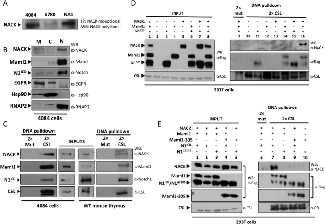
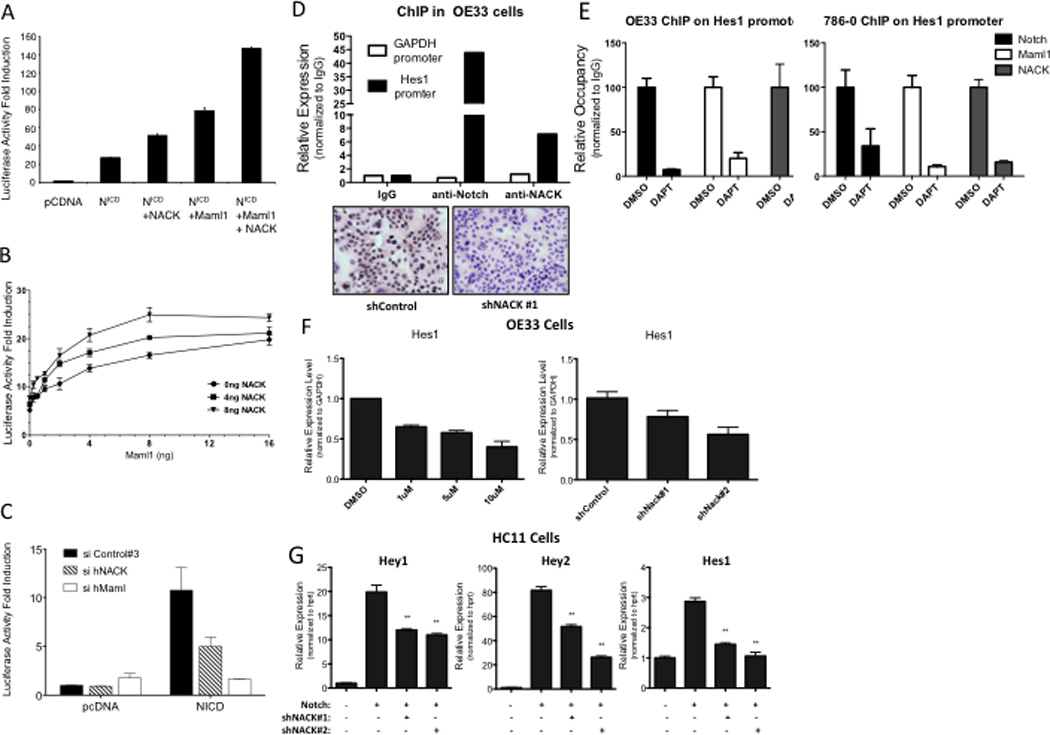
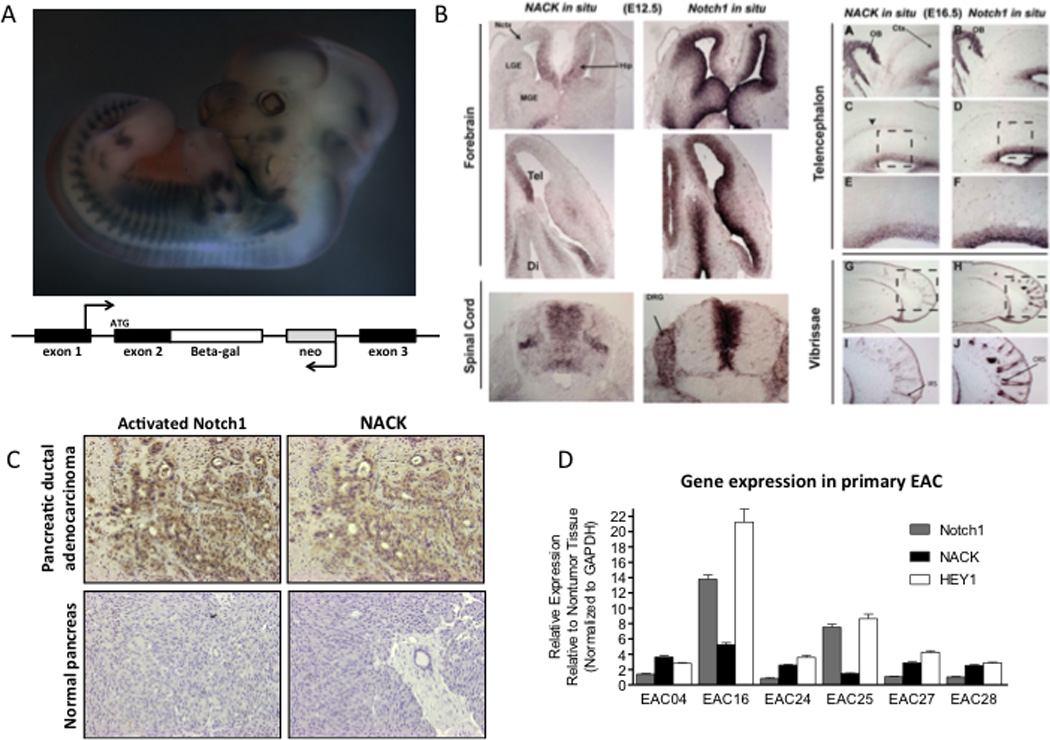
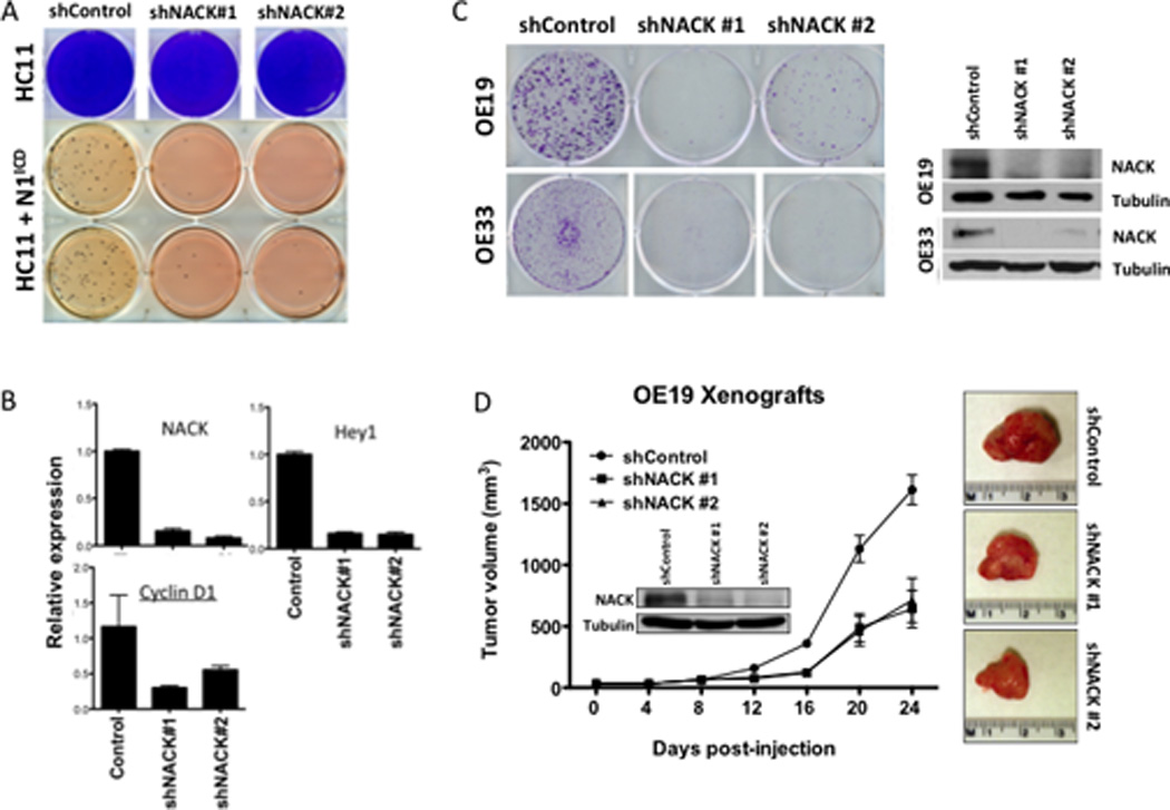
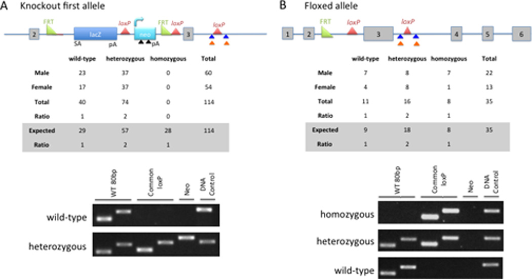
The Notch signaling pathway governs many distinct cellular processes by regulating transcriptional programs. The transcriptional response initiated by Notch is highly cell context dependent, indicating that multiple factors influence Notch target gene selection and activity. However, the mechanism by which Notch drives target gene transcription is not well understood. Herein, we identify and characterize a novel Notch-interacting protein, Notch activation complex kinase (NACK), which acts as a Notch transcriptional coactivator. We show that NACK associates with the Notch transcriptional activation complex on DNA, mediates Notch transcriptional activity, and is required for Notch-mediated tumorigenesis. We demonstrate that Notch1 and NACK are coexpressed during mouse development and that homozygous loss of NACK is embryonic lethal. Finally, we show that NACK is also a Notch target gene, establishing a feed-forward loop. Thus, our data indicate that NACK is a key component of the Notch transcriptional complex and is an essential regulator of Notch-mediated tumorigenesis and development.


Conflict of interest statement

The authors disclose no potential conflicts of interest.

Figures

Figure 1
Figure 1. NACK forms a complex with N1ICD and Maml1
A. IP/western analysis of lysates from T-ALL cell lines. 4084 – Notch-induced lymphoma; 6780 – Myc-induced lymphoma; NA-1 – Ikaros-null lymphoma. B. Western analysis of subcellular fractions of 4084 cells. M – membrane; C – cytoplasm; N – nuclear. C. DNA pull-down and western analysis of nuclear lysates from 4084 cells and wt mouse thymocytes using beads conjugated to 2× CSL binding DNA or 2× mutant CSL binding DNA (2× mut). Input lanes represent 5% of total nuclear lysate. D. DNA pull-down and western analysis of lysates from 293T cells transfected with different combinations of N1ICD, Maml1, and NACK. Input lanes represent 10% of total lysate. E. DNA pull-down and western analysis of lysates from 293T cells transfected with different combinations of N1ICD, N1Δ2105, Maml1, Maml1–305, and NACK. Input lanes represent 10% of total lysate.
Figure 2
Figure 2. NACK is a co-activator of Notch signaling
A–C. 8× CSL luciferase reporter assays in H1299 cells transfected with different combinations of plasmids. Experiments were performed in triplicate and bars represent mean (SEM). A. Histogram of luciferase activity. B. Titration curve showing luciferase activity with different quantities of Maml1 and/or NACK over a fixed quantity of N1ICD (2 ng). C. Histogram of luciferase activity after siRNA-mediated knockdown of Maml or NACK. D. ChIP of Notch and NACK on the Hes1 promoter in OE33 cells. IHC validates specificity of the α-NACK antibody. E. ChIP of Notch, Maml, and NACK on the Hes1 promoter in OE33 and 786-0 cells after treatment with DAPT. F. Hes1 expression in OE33 cells treated with DMSO or DAPT, or infected with shRNA against NACK. Bars represent mean (SEM) of 3 samples. **p<0.01 versus shControl. G. Expression of Notch target genes in HC11 cells infected with N1ICD and shRNA against NACK. Bars represent mean (SEM) of 3 samples. **p<0.01 versus Notch.
Figure 3
Figure 3. NACK is a transcriptional target of Notch signaling
A. mRNA expression in mouse lymphoma cells expressing exogenous N1ICD or wt mouse thymocytes. B. mRNA expression in MEFs infected with N1ICD. C. NACK expression in HC11 cells infected with N1ICD, N2ICD, N3ICD, or N4ICD. Histogram shows mean (SEM). D. Schematic representation showing architecture of the NACK promoter. Numbers denote genomic sequence from − to + with respect to the translation initiation codon ATG. TSS, transcription start site. E. ChIP of Notch on the NACK promoter in OE33 cells.
Figure 4
Figure 4. NACK is co-expressed with Notch during development and in tumors
A. Upper panel, β-galactosidase staining of transgenic knock-in mouse E16.5 embryo. Bottom panel, schematic representation showing knock-in construct. B. In situ hybridization of NACK and Notch1 in mouse central nervous system during development. C. IHC of activated N1ICD and NACK in pancreatic ductal adenocarcinoma and normal pancreas (representative samples). D. Expression of Notch1, NACK, and Hey1 in esophageal adenocarcinoma and adjacent esophageal mucosa. Histogram shows mean (SEM).
Fig. 5
Fig. 5. NACK is required for Notch-mediated transformation and tumorigenesis
A. Upper panel, colony growth of HC11 cells infected with shRNA against NACK. Lower panel, growth of HC11 cells infected with N1ICD and shRNA against NACK in soft agar. B. Expression of NACK, Hey1, and Cyclin D1 in HC11 cells infected with shRNA against NACK. Bars represent mean (SEM) of fold-change relative to control shRNA. C. Colony formation in OE19 and OE33 cells infected with shRNA against NACK. NACK knockdown was verified by western blot. D. Xenograft formation from OE19 cells infected with shRNA against NACK. Tumor volume was measured weekly and error bars indicate SEM. n=8 per group. At 24 d post-injection, tumors were harvested and representative images are shown from each group.
Figure 6
Figure 6. NACK knockout is embryonic lethal
A. Upper panel, schematic of knockout first allele. Colored arrows mark the locations of the primers used for genotyping: black triangle, Neo primers; blue triangle, common loxP primers; orange triangle, WT 80bp primers. Middle panel, summary of live births achieved from mating two NACKKOF/+ mice. Bottom panel, genotyping results from wt and heterozygous pups. B. Top panel, schematic of floxed allele resulting from FLPe recombination. Middle panel, summary of live births achieved from mating two NACKflox/+ mice. Bottom panel, representative genotyping results from wt, heterozygous, and homozygous pups.

References

    1. Weng AP, Ferrando AA, Lee W, Morris JP, Silverman LB, Sanchez-Irizarry C, et al. Activating mutations of NOTCH1 in human T cell acute lymphoblastic leukemia. Science. 2004;306:269–271. - PubMed
    1. Ranganathan P, Weaver KL, Capobianco AJ. Notch signalling in solid tumours: a little bit of everything but not all the time. Nat Rev Cancer. 2011;11:338–351. - PubMed
    1. Miyamoto Y, Maitra A, Ghosh B, Zechner U, Argani P, Iacobuzio-Donahue CA, et al. Notch mediates TGF alpha-induced changes in epithelial differentiation during pancreatic tumorigenesis. Cancer Cell. 2003;3:565–576. - PubMed
    1. Reedijk M, Odorcic S, Chang L, Zhang H, Miller N, McCready DR, et al. High-level coexpression of JAG1 and NOTCH1 is observed in human breast cancer and is associated with poor overall survival. Cancer Res. 2005;65:8530–8537. - PubMed
    1. Artavanis-Tsakonas S, Rand MD, Lake RJ. Notch signaling: cell fate control and signal integration in development. Science. 1999;284:770–776. - PubMed

Publication types