. 2014 Sep 1;193(5):2531-7.
doi: 10.4049/jimmunol.1400918. Epub 2014 Jul 18.

Inducible nitric oxide synthase in neutrophils and endothelium contributes to ischemic brain injury in mice

Affiliations

Inducible nitric oxide synthase in neutrophils and endothelium contributes to ischemic brain injury in mice

Lidia Garcia-Bonilla et al. J Immunol. .

Abstract

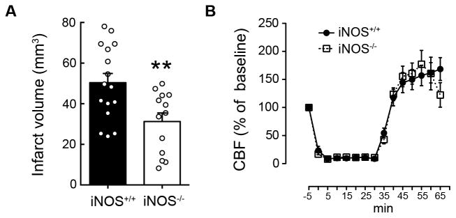
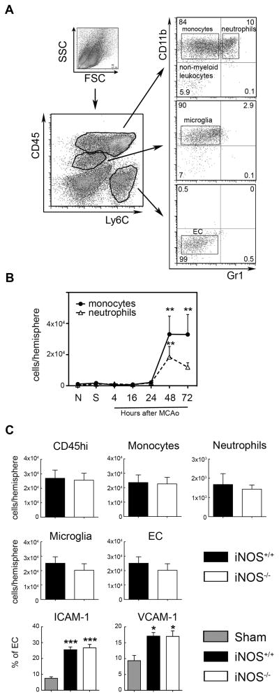
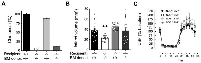
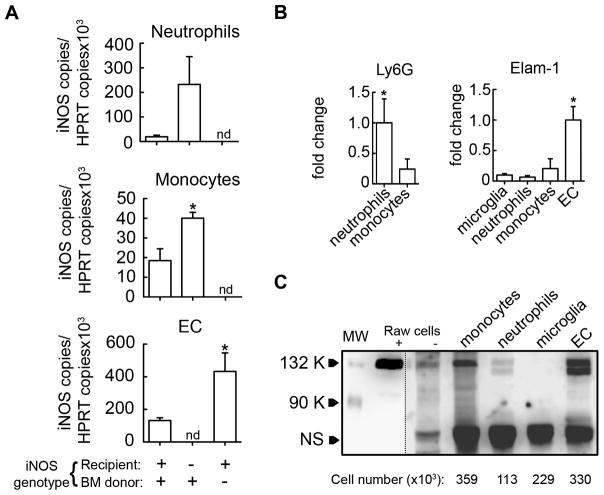
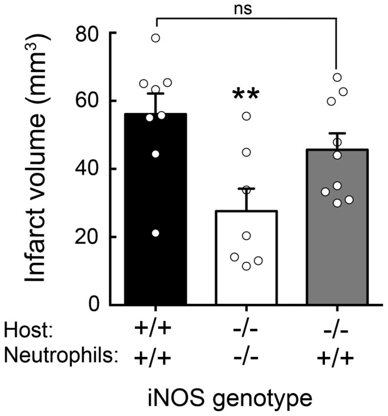
NO produced by inducible NO synthase (iNOS) contributes to ischemic brain injury, but the cell types expressing iNOS and mediating tissue damage have not been elucidated. To examine the relative contribution of iNOS in resident brain cells and peripheral leukocytes infiltrating the ischemic brain, we used bone marrow (BM) chimeric mice in which the middle cerebral artery was occluded and infarct volume was determined 3 d later. iNOS(-/-) mice engrafted with iNOS(+/+) BM exhibited larger infarcts (44 ± 2 mm(3); n = 13; mean ± SE) compared with autologous transplanted iNOS(-/-) mice (24 ± 3 mm(3); n = 10; p < 0.01), implicating blood-borne leukocytes in the damage. Furthermore, iNOS(+/+) mice transplanted with iNOS(-/-) BM had large infarcts (39 ± 6 mm(3); n = 13), similar to those of autologous transplanted iNOS(+/+) mice (39 ± 4 mm(3); n = 14), indicating the resident brain cells also play a role. Flow cytometry and cell sorting revealed that iNOS is highly expressed in neutrophils and endothelium but not microglia. Surprisingly, postischemic iNOS expression was enhanced in the endothelium of iNOS(+/+) mice transplanted with iNOS(-/-) BM and in leukocytes of iNOS(-/-) mice with iNOS(+/+) BM, suggesting that endothelial iNOS suppresses iNOS expression in leukocytes and vice versa. To provide independent evidence that neutrophils mediate brain injury, neutrophils were isolated and transferred to mice 24 h after stroke. Consistent with the result in chimeric mice, transfer of iNOS(+/+), but not iNOS(-/-), neutrophils into iNOS(-/-) mice increased infarct volume. The findings establish that iNOS in both neutrophils and endothelium mediates tissue damage and identify these cell types as putative therapeutic targets for stroke injury.


Figures

Figure 1
Figure 1. Ischemic injury is attenuated in iNOS−/− mice
A. iNOS−/− mice have smaller infarcts than iNOS+/+ 72 hours after MCAo; values are mean±SE, n=13–16/group. **p<0.01. B. Cerebral blood flow (CBF) changes during ischemia and reperfusion were similar in both groups (mean±SE, n=13–16/group).
Figure 2
Figure 2. Brain leukocyte infiltration after MCAo is similar in both iNOS+/+ and iNOS−/− mice
A. Gating strategy and flow cytometric analysis of isolated brain cells stained for CD45, Ly6C, CD11b and Gr1 cell surface markers. Four different populations were identified: CD45hi/CD11bhi/Gr1lo(monocytes), CD45hi/CD11bhi/Gr1hi (neutrophils), CD45int/CD11bhi/Gr1lo/Ly6Clo(microglia) and CD45lo/CD11blo/Gr1lo/Ly6Chi (endothelial cells, EC). B. Time course of infiltrating leukocytes in the ischemic brain of iNOS+/+ mice showed an increase of both monocytes and neutrophils as early as 48 hours after MCAo. Values are mean±SE, n=5–7/group. **P<0.01, vs. naïve (N) and sham (S) groups. C. Total infiltrating leukocytes (CD45hi), monocytes and neutrophils showed similar levels in either iNOS+/+ or iNOS−/− 48 hours after MCAo. No changes were also detected in the number of microglia or EC cells. In addition, the % of EC expressing ICAM-1 and VCAM-1 was similar in both groups. Values are mean±SE, n=6/group. ***p<0.001, *p<0.5 vs. iNOS+/+ sham group.
Figure 3
Figure 3. iNOS expression either in blood-borne leukocytes or resident brain cells reconstitute infarct volume after MCAo
A. Percentage of chimerism in blood leukocytes assayed by genomic qRT-PCR; nd, not detected. Values are mean±SE. B. Transplanted wild type mice (iNOS+/+BM+/+, n=14) had larger infarcts than transplanted iNOS null mice (iNOS−/−BM−/−, n=10) 72 hours after MCAo. iNOS−/− mice expressing iNOS only in peripheral leukocytes (iNOS−/−BM+/+, n=13) or iNOS+/+ mice expressing iNOS only in resident brain cells (iNOS+/+BM−/−; n=13) had similar infarcts to iNOS+/+BM+/+. Values are mean±SE. **p<0.01, vs. iNOS+/+BM+/+. C. No statistical significances in cerebral blood flow (CBF) during ischemia and reperfusion were observed among groups.
Figure 4
Figure 4. iNOS is induced either in brain infiltrating cells or endothelium
Flow cytometric sorting was performed to purify neutrophils, monocytes, microglia and endothelial cells (EC) after staining for CD45, Ly6C, CD11b and Gr1 cell surface markers as described in figure 2. A. Flow-sorted cell fractions from ischemic brains of chimera mice were processed for qRT-PCR to quantify iNOS mRNA expression. In iNOS+/+BM+/+ mice, iNOS mRNA was detected in all cell populations except for microglia (not shown). In iNOS−/− mice transplanted with WT BM cells (iNOS−/−BM+/+), iNOS mRNA was detected in both neutrophils and monocytes, while iNOS mRNA was detected in EC from iNOS+/+ mice transplanted with iNOS−/− BM (iNOS+/+BM−/−). Values are mean±SE, n=3 pool/group, 4 samples pooled; iNOS mRNA expression in chimeric mice vs. iNOS+/+BM+/+ mice, *p<0.05, nd, not detected. B. Ly6G and Elam-1 mRNA was assessed by qRT-PCR in cell fractions to verify purity of separated cell populations. Values are mean±SE, n=3 pool/group, 4 samples pooled. C. iNOS protein levels in neutrophils, monocytes, microglia and endothelial cells (EC) purified by flow cytometric sorting (n=12 pooled mice/cell fraction). The number of cells that corresponds to the amount of lysate loaded into each lane is indicated for each cell population. LPS/IFNγ stimulated (+) or un-stimulated (−) Raw264.7 cells were used as a positive control for iNOS expression. The dash line indicates different exposure time for chemiluminescent detection on the left (short) and right (long) of the blot. Molecular weight, (MW). Non-specific band (NS) served as a loading control.
Figure 5
Figure 5. Adoptive transfer of iNOS+/+ neutrophils worsens ischemic injury in iNOS−/− mice
Adoptive transfer of iNOS+/+ (5×10⁵ cells) but not iNOS−/− neutrophils into iNOS−/− mice 24 hours after MCAO increased infarct volume at 72 hours; values are mean±SE, n=9–8 group. **p<0.05.

References

    1. Iadecola C, Anrather J. The immunology of stroke: from mechanisms to translation. Nat Med. 2011;17:796–808. - PMC - PubMed
    1. Go AS, Mozaffarian D, Roger VL, Benjamin EJ, Berry JD, Blaha MJ, Dai S, Ford ES, Fox CS, Franco S, Fullerton HJ, Gillespie C, Hailpern SM, Heit JA, Howard VJ, Huffman MD, Judd SE, Kissela BM, Kittner SJ, Lackland DT, Lichtman JH, Lisabeth LD, Mackey RH, Magid DJ, Marcus GM, Marelli A, Matchar DB, McGuire DK, Mohler ER, Moy CS, Mussolino ME, Neumar RW, Nichol G, Pandey DK, Paynter NP, Reeves MJ, Sorlie PD, Stein J, Towfighi A, Turan TN, Virani SS, Wong ND, Woo D, Turner MB American Heart Association Statistics Committee and Stroke Statistics Subcommittee. . Heart disease and stroke statistics--2014 update: a report from the American Heart Association. Circulation. 2014;129:e28–e292. - PMC - PubMed
    1. Fonarow GC, Smith EE, Saver JL, Reeves MJ, Bhatt DL, Grau-Sepulveda MV, Olson DM, Hernandez AF, Peterson ED, Schwamm LH. Timeliness of Tissue-Type Plasminogen Activator Therapy in Acute Ischemic Stroke: Patient Characteristics, Hospital Factors, and Outcomes Associated With Door-to-Needle Times Within 60 Minutes. Circulation. 2011;123:750–758. - PubMed
    1. Shichita T, Ago T, Kamouchi M, Kitazono T, Yoshimura A, Ooboshi H. Novel therapeutic strategies targeting innate immune responses and early inflammation after stroke. J Neurochem. 2012;123:29–38. - PubMed
    1. Moskowitz MA, Lo EH, Iadecola C. The science of stroke: mechanisms in search of treatments. Neuron. 2010;67:181–198. - PMC - PubMed

Publication types

MeSH terms