Ultrasonographic diagnosis of round ligament varicosities mimicking inguinal hernia: report of two cases with literature review
- PMID: 25038812
- PMCID: PMC4104952
- DOI: 10.14366/usg.14006
Ultrasonographic diagnosis of round ligament varicosities mimicking inguinal hernia: report of two cases with literature review
Abstract
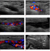
Round ligament varicosities are rare, and the mass mimics an inguinal hernia. Round ligament varicosities should be considered in the differential diagnosis of a groin swelling in a female, especially during pregnancy. The diagnosis of round ligament varicosities can be established on grayscale and color Doppler ultrasonography. We report two cases of round ligament varicosities in a 33-year-old non pregnant woman and a 28-year-old pregnant woman, and these patients were diagnosed using ultrasonography. We also reviewed the literature on round ligament varicosities including the present cases. Ultrasonography is diagnostic and can prevent unnecessary surgical intervention and associated morbidity.
Conflict of interest statement
No potential conflict of interest relevant to this article was reported
Figures


References
-
- Cheng D, Lam H, Lam C. Round ligament varices in pregnancy mimicking inguinal hernia: an ultrasound diagnosis. Ultrasound Obstet Gynecol. 1997;9:198–199. - PubMed
-
- Smith P, Heimer G, Norgren A, Ulmsten U. The round ligament: a target organ for steroid hormones. Gynecol Endocrinol. 1993;7:97–100. - PubMed
-
- Ijpma FF, Boddeus KM, de Haan HH, van Geldere D. Bilateral round ligament varicosities mimicking inguinal hernia during pregnancy. Hernia. 2009;13:85–88. - PubMed
Publication types
LinkOut - more resources
Full Text Sources
Other Literature Sources

