Review
. 2014 Sep;281(18):4085-96.
doi: 10.1111/febs.12936. Epub 2014 Aug 11.

From SARS to MERS: crystallographic studies on coronaviral proteases enable antiviral drug design

Affiliations
Review

From SARS to MERS: crystallographic studies on coronaviral proteases enable antiviral drug design

Rolf Hilgenfeld. FEBS J. 2014 Sep.

Abstract

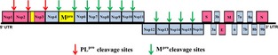
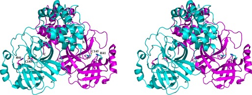
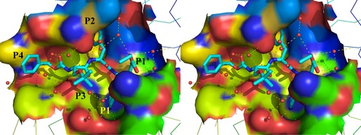
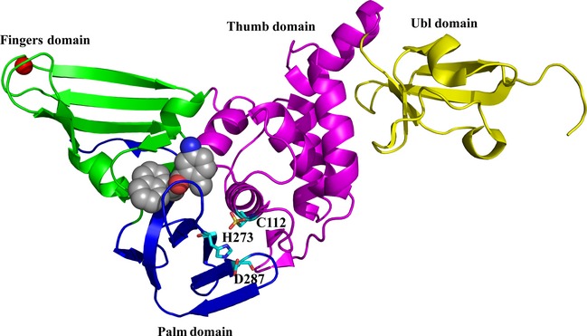
This review focuses on the important contributions that macromolecular crystallography has made over the past 12 years to elucidating structures and mechanisms of the essential proteases of coronaviruses, the main protease (M(pro) ) and the papain-like protease (PL(pro) ). The role of X-ray crystallography in structure-assisted drug discovery against these targets is discussed. Aspects dealt with in this review include the emergence of the SARS coronavirus in 2002-2003 and of the MERS coronavirus 10 years later and the origins of these viruses. The crystal structure of the free SARS coronavirus M(pro) and its dependence on pH is discussed, as are efforts to design inhibitors on the basis of these structures. The mechanism of maturation of the enzyme from the viral polyprotein is still a matter of debate. The crystal structure of the SARS coronavirus PL(pro) and its complex with ubiquitin is also discussed, as is its orthologue from MERS coronavirus. Efforts at predictive structure-based inhibitor development for bat coronavirus M(pro) s to increase the preparedness against zoonotic transmission to man are described as well. The paper closes with a brief discussion of structure-based discovery of antivirals in an academic setting.

Keywords: 3C-like protease; Middle East respiratory syndrome; autoprocessing; bat coronaviruses; high-throughput screening; main protease; papain-like protease; protease maturation; severe acute respiratory syndrome; structure-based inhibitor design.


Figures

Figure 1
Figure 1
Schematic presentation of the genome of the SARS coronavirus. Occupying two‐thirds of the genome from the 5′ end, open‐reading frame 1 (ORF1) encodes two large polyproteins, pp1a and, through ribosomal frameshifting during translation, pp1ab. These polyproteins are processed into mature Nsps by the two proteases discussed here (indicated in yellow). The main protease (Mpro, also called 3C‐like protease, 3CL pro) is Nsp5, whereas the papain‐like protease (PL pro) is a part of Nsp3. The PL pro performs three cleavage reactions (red arrows) to release Nsp1, Nsp2 and Nsp3 (red), whereas the Mpro cleaves the polyprotein at 11 sites (cyan arrows) to release Nsp4–Nsp16 (cyan). The 3′‐terminal third of the genome codes for structural and accessory proteins.
Figure 2
Figure 2
Stereo presentation of the structure of the SARS‐CoV Mpro dimer 36. The catalytic dyads of each subunit (Cys145…His41) are indicated, as are the N‐ and C‐termini. Note that the N‐terminus of the cyan polypeptide chain is located close to the substrate‐binding site of the purple subunit.
Figure 3
Figure 3
Stereo illustration of the Michael acceptor compound SG85 (Cbz–(tBu–O–)Ser–Phe–GlnLactam–CH=CH–CO–O–Et; 59) bound to the substrate‐binding site of the SARS‐CoV Mpro (Zhu et al., unpublished; PDB code http://www.rcsb.org/pdb/search/structidSearch.do?structureId=3TNT). The inhibitor is shown in cyan (for carbon), blue (for nitrogen) and red (for oxygen). Hydrogen bonds are indicated by dashed lines and water molecules by small red spheres. The P4–P1’ side‐chains of the inhibitor are labeled. The P1 side‐chain is buried in the S1 pocket and only the tip of its lactam moiety is visible in this illustration.
Figure 4
Figure 4
Structure of the SARS‐CoV PL pro in complex with the non‐peptidic inhibitor GRL0617 78. The domains of the enzyme are indicated and colored as follows: yellow, ubiquitin‐like (Ubl) domain; pink, thumb domain; blue, palm domain; cyan, fingers domain. The zinc ion bound at the tip of the fingers domain is colored red. The inhibitor (space‐filling presentation, with carbon in grey, nitrogen in blue and oxygen in red) binds to the S3 and S4 sites, far from the catalytic triad, C112–H273–D287 (cyan sticks).

References

    1. Hilgenfeld R & Peiris JSM (2013) From SARS to MERS: 10 years of research on highly pathogenic human coronaviruses. Antiviral Res 100, 286–295. - PMC - PubMed
    1. Cheng VC, Chan JF, To KK & Yuen KY (2013) Clinical management and infection control of SARS: lessons learned. Antiviral Res 100, 407–419. - PMC - PubMed
    1. Graham RL, Donaldson EF & Baric RS (2013) A decade after SARS: strategies for controlling emerging coronaviruses. Nat Rev Microbiol 11, 836–848. - PMC - PubMed
    1. Koplan JP, Butler‐Jones D, Tsang T & Yu W (2013) Public health lessons from severe acute respiratory syndrome a decade later. Emerg Infect Dis 19, 861–863. - PMC - PubMed
    1. Peiris JS, Lai ST, Poon LL, Guan Y, Yam LY, Lim W, Nicholls J, Yee WK, Yan WW, Cheung MT et al, SARS Study Group (2003) Coronavirus as a possible cause of severe acute respiratory syndrome. Lancet 361, 1319–1325. - PMC - PubMed

Publication types