. 2014 Aug 12;53(31):5131-9.
doi: 10.1021/bi500593e. Epub 2014 Jul 28.

The structure and specificity of the type III secretion system effector NleC suggest a DNA mimicry mechanism of substrate recognition

Affiliations

The structure and specificity of the type III secretion system effector NleC suggest a DNA mimicry mechanism of substrate recognition

Michelle Marian Turco et al. Biochemistry. .

Abstract

Many pathogenic bacteria utilize the type III secretion system (T3SS) to translocate effector proteins directly into host cells, facilitating colonization. In enterohemmorhagic Escherichia coli (EHEC), a subset of T3SS effectors is essential for suppression of the inflammatory response in hosts, including humans. Identified as a zinc protease that cleaves NF-κB transcription factors, NleC is one such effector. Here, we investigate NleC substrate specificity, showing that four residues around the cleavage site in the DNA-binding loop of the NF-κB subunit RelA strongly influence the cleavage rate. Class I NF-κB subunit p50 is cleaved at a reduced rate consistent with conservation of only three of these four residues. However, peptides containing 10 residues on each side of the scissile bond were not efficiently cleaved by NleC, indicating that elements distal from the cleavage site are also important for substrate recognition. We present the crystal structure of NleC and show that it mimics DNA structurally and electrostatically. Consistent with this model, mutation of phosphate-mimicking residues in NleC reduces the level of RelA cleavage. We propose that global recognition of NF-κB subunits by DNA mimicry combined with a high sequence selectivity for the cleavage site results in exquisite NleC substrate specificity. The structure also shows that despite undetectable similarity of its sequence to those of other Zn(2+) proteases beyond its conserved HExxH Zn(2+)-binding motif, NleC is a member of the Zincin protease superfamily, albeit divergent from its structural homologues. In particular, NleC displays a modified Ψ-loop motif that may be important for folding and refolding requirements implicit in T3SS translocation.


Figures

Figure 1
Figure 1
Structure of NleC. (A) Structure of NleC with the active site highlighted. The structure of NleC is shown in standard Zincin coloring as described in Gomis-Ruth et al., with helices colored yellow, strands aqua, and loops gray. The active site residues are shown in magenta stick format. The inset shows a close-up of the active site, including the distances between the zinc and coordinating atoms. (B) Modified Ψ-loop β-sheet motif in NleC. The NleC β-sheet is shown in cartoon and stick format. Red dotted lines represent hydrogen bonds between the strands and illustrate the standard β-sheet interactions between all three strands and the outer two strands after the middle strand exits the sheet.
Figure 2
Figure 2
NF-κB proteolysis by NleC. (A) Cleavage of NF-κB subunits by NleC. NF-κB subunits (20 μM), p50, RelA, and RelB, were incubated with 20 nM NleC for 10 min before the reaction was quenched by the addition of SDS–PAGE sample buffer and boiling for 2 min. Results were analyzed by SDS–PAGE. The bands were quantified in ImageJ for visualization in the bar graph. (B) DNA-binding loop in Rel homology domains. Sequence alignment of the residues in the DNA-binding loop of NF-κB subunits RelA, RelB, and p50 as well as transcription factor NFATc2. The scissile bond is shown as a red dotted line. In green are the residues whose mutation to alanine strongly affects NleC cleavage. Light green delineates the arginine that has an intermediate effect on NleC cleavage upon being mutated to alanine, and the proline that is in the same position in p50. The sequence of the NFATc2 DNA-binding loop is shown for reference. (C) DNA-binding loop of RelA containing the scissile bond. Four residues to either side of the scissile bond between cysteine 38 and glutamate 39 are colored green in the DNA-binding domain structure of RelA (PDB entry 2RAM). (D) Cleavage of RelA mutants by NleC. RelA (20 μM), wild type or alanine mutants, was incubated with 20 nM NleC for 10 min before the proteolysis reaction was quenched via addition of SDS–PAGE sample buffer and boiling for 2 min. The results were analyzed by SDS–PAGE and the bands quantified in ImageJ for visualization in bar graph form.
Figure 3
Figure 3
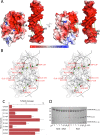
DNA mimicry in NleC. (A) Electrostatic surface potentials of NleC and DNA. Electrostatic surface potentials calculated with APBS are shown mapped to the van der Waals surfaces of NleC and DNA. Green bars illustrate the distances across the major groove of RelA-bound DNA and the active site groove of NleC as measured with Pymol. The right panel is rotated 90° from the left. (B) Superposition of NleC and DNA. Stereoview of the cartoon and semitransparent surface representation of NleC superimposed on a cartoon representation of DNA aligning negatively charged residues in NleC with the phosphate backbone of DNA. Glutamic acid and aspartic acid residues on the active site face of NleC that overlay with DNA phosphates are colored red, and the carboxyl carbon is depicted as a sphere. The carboxyl carbons of other nearby negatively charged residues are depicted as salmon-colored spheres. The phosphates in DNA that are contacted by RelA in the majority of DNA–RelA crystal structures (PDB entries 1RAM, 2RAM, 1LE5, 2I9T, and 3GUT) are colored dark green, with other phosphates that overlay with negatively charged NleC residues colored light green. The superposition was done manually to maximize shape similarity and charge correspondence between NleC and DNA. (C) Alanine scanning mutagenesis of NleC negative charges. RelA (20 μM) was incubated with 20 nM wild-type NleC or 20 nM NleC mutant for 10 min before the reaction was quenched with SDS–PAGE sample buffer and boiling for 2 min. The control reaction mixture contained no NleC. The wild-type NleC reaction was repeated with three biological replicates, which were each tested three times. All NleC mutant reactions are the result of one biological replicate, repeated three times with different dilutions. The results were analyzed by SDS–PAGE and the bands quantified in ImageJ for analysis (Figure S5 of the Supporting Information). The error bars indicate the standard error = standard deviation. (D) Effect of DNA on NleC proteolysis of RelA. After incubation of 20 μM RelA with (left) or without (right) a 1:1 molar ratio of palindromic DNA encoding the RelA-binding site, 20 nM NleC was added and reaction time points were taken. Reactions were quenched via the addition of SDS–PAGE sample buffer and boiling for 5 min and the products analyzed by SDS–PAGE.

References

    1. Baron C.; Coombes B. (2007) Targeting bacterial secretion systems: Benefits of disarmament in the microcosm. Infect. Disord.: Drug Targets 7, 19–27. - PubMed
    1. Wong A. R. C.; Pearson J. S.; Bright M. D.; Munera D.; Robinson K. S.; Lee S. F.; Frankel G.; Hartland E. L. (2011) Enteropathogenic and enterohaemorrhagic Escherichia coli: Even more subversive elements. Mol. Microbiol. 80, 1420–1438. - PubMed
    1. Donnenberg M. S.; Kaper J. B. (1992) Enteropathogenic Escherichia coli. Infect. Immun. 60, 3953. - PMC - PubMed
    1. McDaniel T. K.; Jarvis K. G.; Donnenberg M. S.; Kaper J. B. (1995) A genetic locus of enterocyte effacement conserved among diverse enterobacterial pathogens. Proc. Natl. Acad. Sci. U.S.A. 92, 1664–1668. - PMC - PubMed
    1. Frankel G.; Phillips A. D.; Rosenshine I.; Dougan G.; Kaper J. B.; Knutton S. (1998) Enteropathogenic and enterohaemorrhagic Escherichia coli: More subversive elements. Mol. Microbiol. 30, 911–921. - PubMed

Publication types

MeSH terms

Associated data