. 2015 Feb;61(2):627-38.
doi: 10.1002/hep.27306. Epub 2015 Jan 5.

CXCR5+ CD4+ T follicular helper cells participate in the pathogenesis of primary biliary cirrhosis

Affiliations

CXCR5+ CD4+ T follicular helper cells participate in the pathogenesis of primary biliary cirrhosis

Lifeng Wang et al. Hepatology. 2015 Feb.

Abstract

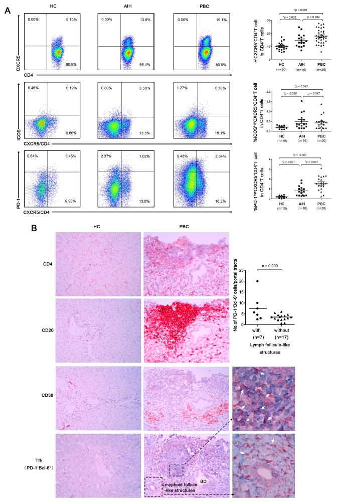
There is increasing interest in the role of T follicular helper (Tfh) cells in autoimmunity from the perspective of both their role in breach of tolerance and their effects on the natural history of disease progression. Indeed, the critical role of Tfh cells in autoimmunity is further highlighted based on their location in the germinal center (GC), a pathogenic hot spot for development of autoreactivity. To address the role of Tfh cells in primary biliary cirrhosis (PBC), we comprehensively evaluated the immunobiology of CXCR5(+) CD4(+) Tfh cells in 69 patients with PBC, including a nested subgroup of 16 autoimmune hepatitis (AIH) and 20 healthy controls (HC), followed for 1 year. We report herein several key observations. First, there was an increased frequency of circulating Tfh cells in patients with PBC compared to AIH (P < 0.05) and HC (P < 0.01). Second, the function of circulating Tfh cells from PBC patients, including interleukin (IL)-21 production (P < 0.05), the ability to promote B-cell maturation, and autoantibody production, were greater than HC. Third, the frequency of these cells was significantly decreased in ursodeoxycholic acid (UDCA) responders compared to UDCA-treated nonresponders, in both cross-sectional (P = 0.023) and longitudinal studies (P = 0.036), respectively. Indeed, similar increases of Tfh cells were noted in liver and spleen.

Conclusion: These results significantly extend our understanding of lymphoid subpopulations in PBC and their relative role in disease expression. Our data also provide a novel biomarker for evaluation of the effectiveness of new therapeutic approaches.


Figures

Fig 1
Fig 1. Increased frequency of follicular helper T (Tfh) cells in primary biliary cirrhosis
(A) Comparison of the frequencies of total circulating Tfh in PBC, AIH and healthy controls (HCs). PBMC from PBC (n = 35) , AIH (n = 16) and HCs (n = 20) were stained with labeled antibodies. Representative expressions of CXCR5+CD4+ (and ICOShigh CXCR5+CD4+ or PD-1high CXCR5+CD4+) versus CD4 expression were detected by flow cytometry. Representative dot plots are shown in the right panel. (B) Intrahepatic double positive Bcl-6 (blue) and PD-1 (red) cells were found around chronic non-suppurative destructive cholangitis (CNSDC) in PBC (n = 24), but not in HCs. The white arrow indicates the Bcl-6+PD-1+ Tfh cells.
Fig 2
Fig 2. Peripheral Tfh cells correlate with disease severity
(A) Higher frequencies of circulating Tfh cells were found in AMA+ PBC (n = 29) than in AMA patients (n = 6), p <0.05. (B) There was a higher frequency of circulating Tfh cells based on disease severity. (C) Circulating Tfh cells were higher in late stages (II, III and IV) compared with early stage (I). Horizontal lines illustrate the median percentiles. P values are shown.
Fig 3
Fig 3. Cytokine production by CXCR5+CD4+ T cells
(A) Representative results reflect cytokine production by CXCR5+CD4+ T cells (upper part). CXCR5+CD4+ T cells release more IL-21, IFN- and IL-17 in PBC (n = 24) than in HCs (n = 10) (lower part) when stimulated with PMA/ionomycin. *: p <0.05; **: p <0.01, respectively. (B) CXCR5+CD4+ T cells produced higher levels of IL-21 than IL-6 and IL-17 compared with CXCR5CD4+ T cells; while, CXCR5CD4+ T cells predominantly produced IFN-γ. PBMCs from 24 patients were analyzed. P values are shown. (C) Correlation between the frequency of CXCR5+CD4+ T cells and IL-21 secretion from 24 PBC patients when stimulated with PMA/ionomycin (p < 0.05).
Fig 4
Fig 4. Increased Tfh cells correlate with plasma cell proportions and affect B cell function in PBC
(A) Percentages of CXCR5+CD4+ T cells correlated with the percentage of CD24CD38++CD27+CD19+ plasma cells in 16 treatment-naïve PBC patients (r = 0.513, p <0.05), but were not correlated with total B cells. (B) Patient's CXCR5+CD4+ T (pTfh) promoted more proliferation and differentiation of allogeneic B cells compared with that of HCs’ CXCR5+CD4+ T (hTfh) and CXCR5+CD4 T cells derived from PBC (pTfh -) or healthy controls (hTfh-). *: p <0.05; **: p <0.01, respectively. (C) CXCR5+CD4+ T fh from PBC (pTfh) promoted higher production of IgG and IgM compared with CXCR5+CD4+ T from HCs (hTfh). Exogenous IL-21 or blockade of IL-21 with soluble rIL-21R-Fc resulted in significantly increased or decreased immunoglobulin production by co-cultured B cells, respectively. Five PBC patients and four HCs were studied. Representative data are from three independent experiments.
Fig 5
Fig 5. Decreased proportion of circulating Tfh cells is detected in UDCA responders
(A) The frequency of CXCR5+CD4+ T cells in UDCA responders (n = 14) was significantly lower than that of UDCA non-responders (n = 14). p < 0.05 (B) Comparison of IL-21 secretion by CXCR5+CD4+ T cells between UDCA responders (n = 12) and non-responders (n = 8). There was a decreased trend of IL-21 production by CXCR5+CD4+ T cells from UDCA responders compared with those from UDCA non-responders. (C) After 1 year of follow-up, there were 10 UDCA responders and 8 non-responders. The percentages of circulating CXCR5+CD4+ T cells were reduced at 12 months compared with entry data in the UDCA responders group. In contrast, no significant changes were observed in the non-responders group during the 1-year follow-up period. P values are shown.

Comment in

References

    1. Kaplan MM, Gershwin ME. Primary biliary cirrhosis. N Engl J Med. 2005;353:1261–1273. - PubMed
    1. Selmi C, Bowlus CL, Gershwin ME, Coppel RL. Primary biliary cirrhosis. Lancet. 2011;377:1600–1609. - PubMed
    1. Kita H, Lian ZX, Van de Water J, He XS, Matsumura S, Kaplan M, Luketic V, et al. Identification of HLA-A2-restricted CD8(+) cytotoxic T cell responses in primary biliary cirrhosis: T cell activation is augmented by immune complexes cross-presented by dendritic cells. J Exp Med. 2002;195:113–123. - PMC - PubMed
    1. Shimoda S, Nakamura M, Ishibashi H, Hayashida K, Niho Y. HLA DRB4 0101-restricted immunodominant T cell autoepitope of pyruvate dehydrogenase complex in primary biliary cirrhosis: evidence of molecular mimicry in human autoimmune diseases. J Exp Med. 1995;181:1835–1845. - PMC - PubMed
    1. Takahashi T, Miura T, Nakamura J, Yamada S, Miura T, Yanagi M, Matsuda Y, et al. Plasma cells and the chronic nonsuppurative destructive cholangitis of primary biliary cirrhosis. Hepatology. 2012;55:846–855. - PMC - PubMed

Publication types