. 2014 Jul 31;511(7511):616-20.
doi: 10.1038/nature13393. Epub 2014 Jun 22.

Targeting transcription regulation in cancer with a covalent CDK7 inhibitor

Affiliations

Targeting transcription regulation in cancer with a covalent CDK7 inhibitor

Nicholas Kwiatkowski et al. Nature. .

Abstract

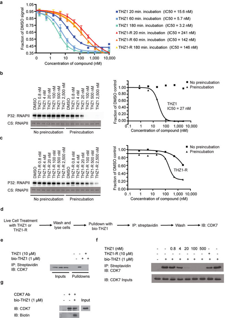
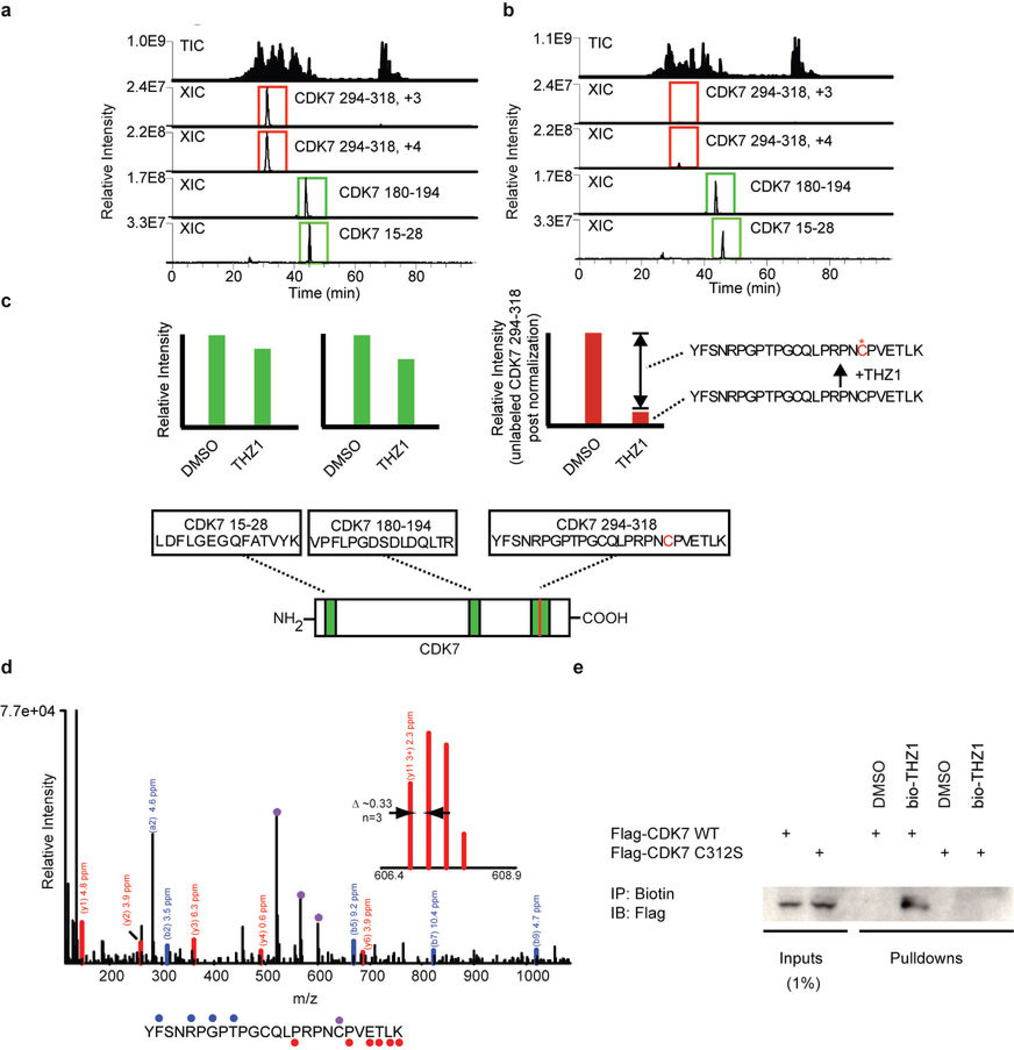
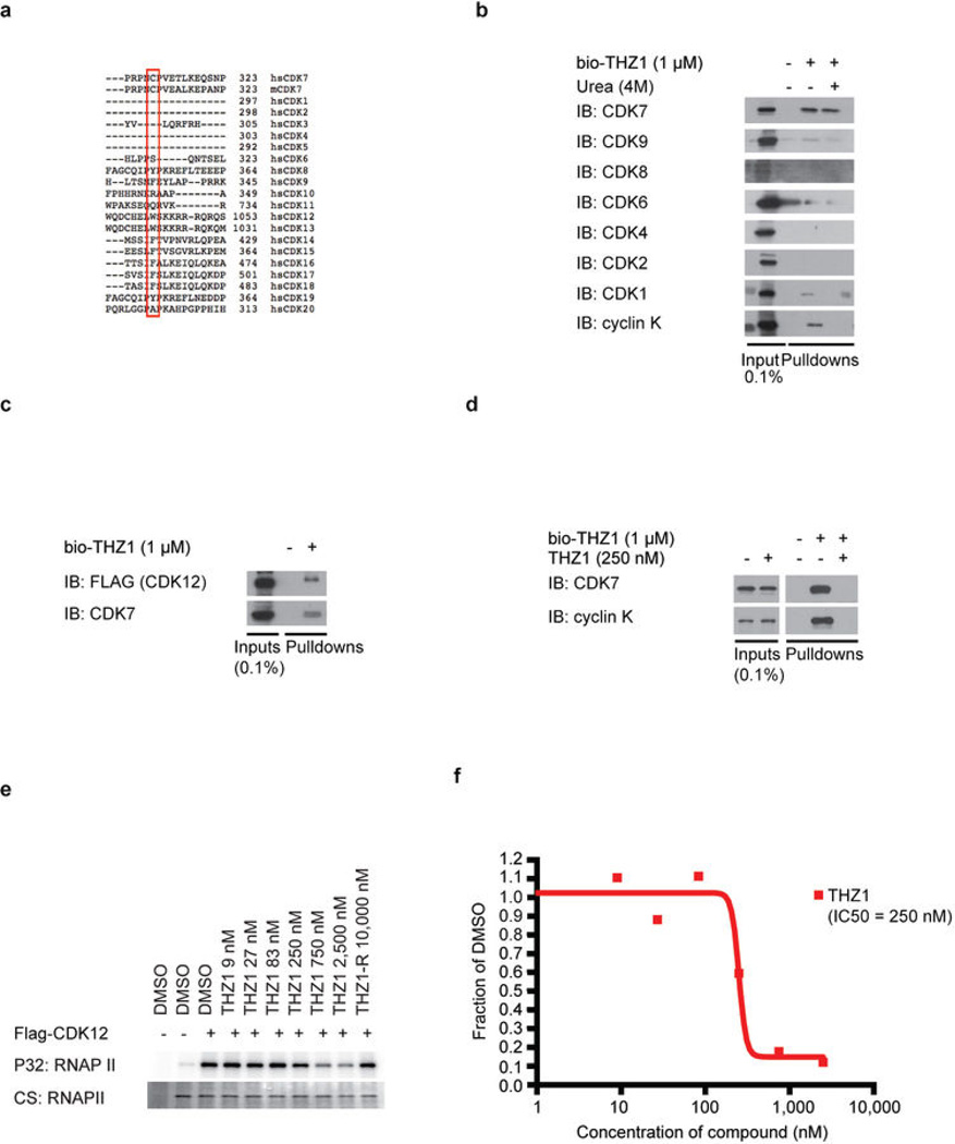
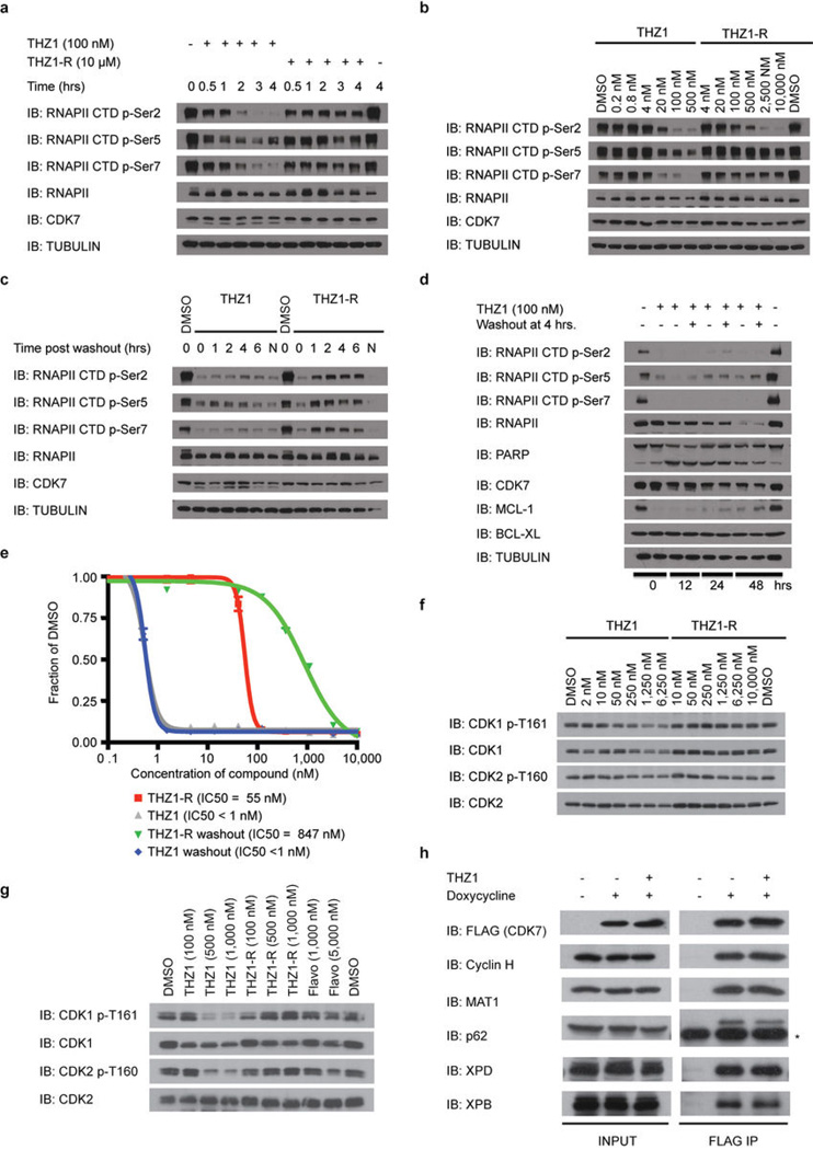
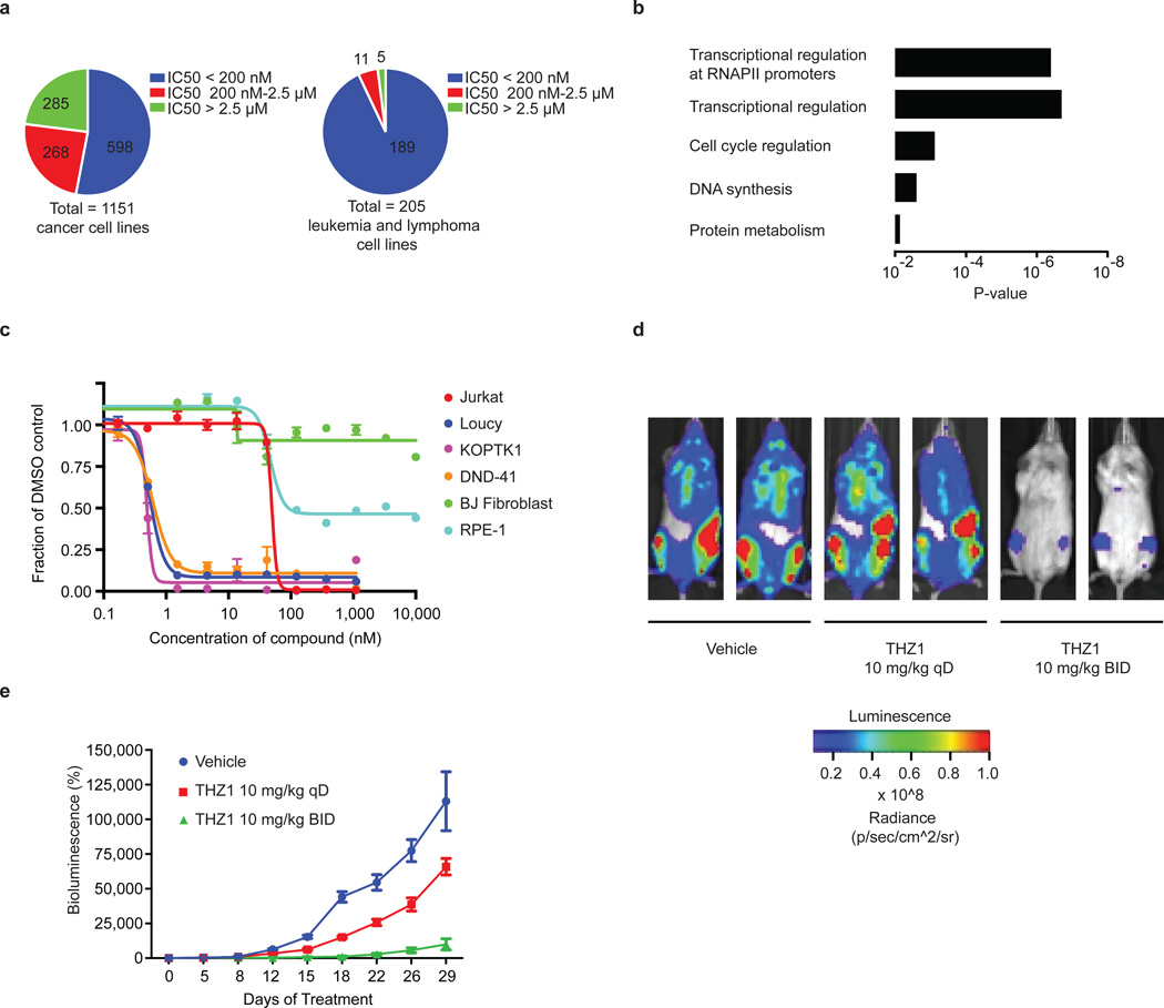
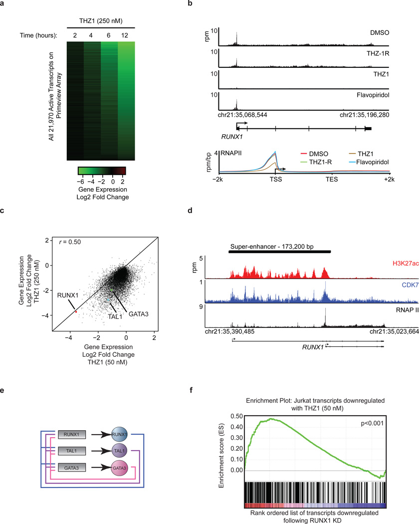
Tumour oncogenes include transcription factors that co-opt the general transcriptional machinery to sustain the oncogenic state, but direct pharmacological inhibition of transcription factors has so far proven difficult. However, the transcriptional machinery contains various enzymatic cofactors that can be targeted for the development of new therapeutic candidates, including cyclin-dependent kinases (CDKs). Here we present the discovery and characterization of a covalent CDK7 inhibitor, THZ1, which has the unprecedented ability to target a remote cysteine residue located outside of the canonical kinase domain, providing an unanticipated means of achieving selectivity for CDK7. Cancer cell-line profiling indicates that a subset of cancer cell lines, including human T-cell acute lymphoblastic leukaemia (T-ALL), have exceptional sensitivity to THZ1. Genome-wide analysis in Jurkat T-ALL cells shows that THZ1 disproportionally affects transcription of RUNX1 and suggests that sensitivity to THZ1 may be due to vulnerability conferred by the RUNX1 super-enhancer and the key role of RUNX1 in the core transcriptional regulatory circuitry of these tumour cells. Pharmacological modulation of CDK7 kinase activity may thus provide an approach to identify and treat tumour types that are dependent on transcription for maintenance of the oncogenic state.


Figures

Extended Data Figure 1
Extended Data Figure 1. THZ1 demonstrates time-dependent inhibition of CDK7 in vitro and covalent binding of intracellular CDK7
a, THZ1 but not THZ1-R shows time-dependent inhibition. LanthaScreen® Eu Kinase Binding assay was conducted at Life Technologies in a time-dependent manner (20, 60, and 180 min.) showing that THZ1 but not THZ1-R shows time-dependent inhibition of CDK7. b and c, Pre-incubation of THZ1 increases CDK7 inhibitory activity in vitro. Recombinant CAK complex was incubated with THZ1 (b) or THZ1-R (c) in dose response format with or without pre-incubation prior to ATP (25 µM) addition. Kinase reaction was then allowed to proceed for 45 minutes at 30°C. d, Workflow of bio-THZ1 pull down competition experiment. e, bio-THZ1 pulls down CDK7 from cellular lysates. Loucy cellular lysates were incubated with bio-THZ1 (1 µM) with or without THZ1 (10 µM) and streptavidin-precipitated proteins were probed for CDK7. IB = immunoblot. f, Free intracellular THZ1 competes in a dose-dependent manner for bio-THZ1 binding to CDK7. Loucy cells were treated with increasing concentrations of THZ1 or with 10 µM THZ1-R for 4 hrs. Cellular lysates were incubated with bio-THZ1 and processed as indicated in a. g, bio-THZ1 labels CDK7 in lysates. Loucy cellular lysates were incubated with bio-THZ1 at 4°C for 12 hrs followed by immunoprecipitation of CDK7 at 4°C for 3 hrs. Precipitated proteins were washed and probed with streptavidin-HRP.
Extended Data Figure 2
Extended Data Figure 2. THZ1 covalently binds CDK7 C312
a and b, Total ion chromatograms (TIC) and extracted ion chromatograms (XIC) for CDK7 peptides recorded during analysis of CAK complexes treated with DMSO (a) or THZ1 (b). c, Efficiency of labeling was estimated to be approximately 85% gauged by the reduction in signal of triply and quadruply charged YFSNRPGPTPGCQLPRPNCPVETLK ions (residues 294–318). The peptides VPFLPGDSDLDQLTR (residues 180–194) and LDFLGEGQFATVYK (residues 15–28) were used for normalization. d, Orbit rap HCD MS/MS spectrum of a quadruply charged CDK7 derived peptide (residues 294–318) labeled by THZ1 at C312. Fragment ions containing the peptide C-terminus (y-type) or N-terminus (b-type), along with the associated mass errors are shown in red and blue, respectively. Fragment ions marked by (*) contain the inhibitor and have the expected heavy isotope contribution from chlorine. The site of labeling was determined to be C312 (as opposed to C305) based on fragment ions observed in additional MS/MS spectra (for example y113+ observed with < 3 ppm mass error by fragmentation of the +6 charged precursor; see inset mass spectrum). e, C312S mutation eliminates THZ1 covalent binding. Cellular lysates from HCT116 cells expressing either FLAG-CDK7 WT or C312S were incubated with bio-THZ1 for 12 hrs at 4°C and then room temperature for 3 hrs to facilitate covalent binding. Precipitated proteins were then probed for the presence of FLAG-tagged CDK7.
Extended Data Figure 3
Extended Data Figure 3. THZ1 inhibits CDK12 but at higher concentrations compared to CDK7
a, Protein sequence alignment of the C-terminal regions of all human (hs) CDKs and mouse (m) CDK7 using Uniprot default settings. Note that the canonical cell cycle CDKs 1,2,4 as well as 5 do not have C-terminal domains that extent to the equivalent position of CDK7 C312 and therefore do not display aligned sequence in this region. b, bio-THZ1 covalently pulls down CDK7 from cellular lysates. Jurkat cellular lysates were incubated with bio-THZ1 (1 µM) at 4°C for 12 hrs and 2 hrs at room temperature. Precipitated proteins were washed with or without urea (4M), here used as a denaturing agent, and probed for the indicated CDKs. c, bio-THZ1 pulls down FLAGCDK12 from lysates. Lysates from 293A cells stably expressing FLAG-tagged WT CDK12 were incubated with bio-THZ1 (1 µM) at 4°C for 12 hrs and 2 hrs at rt. Immunoprecipitated proteins were probed with FLAG antibody to recognize CDK12 or with CDK7 antibody. d, bio-THZ1 pulls down cyclin K from cellular lysates. Jurkat cellular lysates were incubated with bio-THZ1 (1 µM) at 4°C for 12 hrs and 2 hrs at rt. Precipitated proteins were probed for the indicated proteins. e, THZ1 inhibits CDK12 in an in vitro kinase assay. 293A cells stably expressing FLAG-tagged WT CDK12 were treated with THZ1 or THZ1-R for 4 hrs. Exogenous CDK12 was immunoprecipitated from cellular lysates using FLAG antibody. Precipitated proteins were washed and subjected to in vitro kinase assays at 30°C for 30 minutes using the large subunit of RNAPII (RPB1) as substrate and 25 µM ATP. CS = coomassie stain. f, Quantitation of in vitro kinase assay conducted in (d).
Extended Data Figure 4
Extended Data Figure 4. THZ1 irreversibly inhibits RNAPII CTD and CAK phosphorylation
a, THZ1 exhibits time-dependent inactivation of intracellular CDK7. Loucy cells were treated with THZ1 or THZ1-R for 0 to 4 hrs. At each time point cells were harvested, lysed, and the cellular lysates were probed with antibodies against the specified proteins. b, THZ1 inhibits RNAPII CTD phosphorylation. Loucy cells were treated with THZ1 or THZ1-R for 4 hrs. Cellular lysates were then probed with antibodies recognizing the Ser-2, Ser-5, and Ser-7 CTD RNAPII phosphoepitopes. c, Loucy cells were treated with THZ1 or THZ1-R for 4 hrs followed by washout of inhibitor-containing medium. Cells were allowed to grow in medium without inhibitor for 0 to 6 hrs. At each time point cells were lysed and the cellular lysates were probed with antibodies against the specified proteins. ‘N’ signifies cells where medium was never washed out. d, Apoptotic signaling is maintained despite washout of THZ1. Loucy cells were treated with THZ1 or THZ1-R for 4 hrs followed by washout of inhibitor-containing medium, at which point cells were allowed to grow in medium with or without inhibitor for 0 to 48 hrs. At each time point cells were lysed and the cellular lysates were probed with antibodies against the specified proteins. e, Anti proliferative effects of THZ1 are impervious to inhibitor washout. Loucy cells were treated with THZ1 or THZ1-R in dose response format for 72 hrs. Anti proliferative effects were determined using cell titer glo analysis. f, THZ1 reduces the T-loop phosphorylation status of CDK1 and CDK2 in Jurkat cells over a 3 hour exposure. Asynchronous cells were treated with increasing concentrations of THZ1 or THZ1-R for 3 hrs. Cellular lysates were then probed with antibodies against the indicated proteins or phosphoproteins. g, THZ1, but not THZ1-R, completely inhibits T-loop phosphorylation of CDK1 and CDK2 following treatment over one cell cycle. Loucy cells were treated with THZ1, THZ1-R, Flavopiridol, or DMSO vehicle at the indicated concentrations for 24 and 14 hrs, respectively (roughly one cell cycle). Cell lysates were harvested and probed with antibodies against the specified proteins or phosphoproteins. h, Hela S3 cells stably expressing FLAG-WT CDK7 were treated with THZ1 (1 µM) or DMSO vehicle for 5 hrs with and without the presence of doxycycline. Proteins were immunoprecipitated using FLAG antibody. Precipitated proteins were probed using the indicated antibodies. * indicates heavy-chain from IgG antibody.
Extended Data Figure 5
Extended Data Figure 5. Mutation of CDK7 Cys-312 to serine rescues Ser-5/7 and partially Ser2RNAPII CTD phosphorylation
a, Expression of C312S rescues Ser-5/7 and partially rescues Ser-2 RNAPII CTD phosphorylation. Hela S3 cells stably carrying a doxycycline-inducible FLAG-C312S CDK7 construct were treated with THZ1 or DMSO for 5 hrs with and without the presence of doxycycline. Cellular lysates were then probed for the indicated proteins. b, Phenotypic rescue is specific to C312S mutation as rescue is not achieved with overexpression of FLAG-WT CDK7. Hela S3 cells stably carrying doxycycline-inducible FLAG-WT and C312S CDK7 constructs (or empty vector) were treated with THZ1 or DMSO for 5 hrs in the presence of doxycycline. c, Expression of C312S largely restores CDK1/2 T-loop phosphorylation. Hela S3 cells stably carrying a doxycycline-inducible FLAG-C312S CDK7 construct were treated with THZ1 or DMSO for 5 hrs with and without the presence of doxycycline. Cellular lysates were then probed for the indicated proteins or phosphoproteins. d, Overexpression of FLAG-CDK7 C312 rescues the expression of a subset of transcripts in Hela S3 cells. Log2 fold change in gene expression in Hela S3 cells expressing FLAG-CDK7 WT (x axis) and FLAG-CDK7 C312S (y axis) following a 4 hr treatment with 500 nM THZ1. e, Gene ontology molecular function analysis of transcripts increased by 1 log2 order or more following expression of FLAG-CDK7 C312S compared to FLAG-CDK7 WT in the presence of 500 nM THZ1.
Extended Data Figure 6
Extended Data Figure 6. THZ1 potently disrupts T-ALL proliferation
a, THZ1, but not THZ1-R, exhibits strong antiproliferative effect against T-ALL cell lines. Cells were treated with THZ1, THZ1-R, or DMSO vehicle for 72 hrs and assessed for antiproliferative effect by Cell Titer Glo analysis. Error bars are +/− SD. b, THZ1 causes cell cycle arrest. Jurkat (top) and Loucy (bottom) T-ALL cells were treated with THZ1 for the indicated time periods. Cell cycle progression was assessed using FACS cell cycle analysis. 2N = G1, 4N = G2. c, Treatment with THZ1 decreases CDK1/2 T loop phosphorylation. Jurkat cells were incubated with THZ1 for the indicated duration of time and lysates were probed for the specified proteins.
Extended Data Figure 7
Extended Data Figure 7. Treatment with THZ1 induces apoptosis in T-ALL cells
a, Representative Annexin V and propidium iodide stainings for Jurkat cells incubated with THZ1 for the indicated amount of time and harvested to determine the percentage of apoptotic and/ or dead cells by Annexin V and propidium iodide staining, respectively. The percentage of cells in each cell population is shown in the four quadrants. b, Treatment with THZ1 induces apoptosis. Quantitation of Annexin V and propidium iodide staining data from a. Experiments were performed in biological triplicates and error bars are +/− SD. c, Representative Annexin V and propidium iodide stainings for Loucy cells incubated with THZ1 for the indicated amount of time and harvested to determine the percentage of apoptotic and/ or dead cells by Annexin V and propidium iodide staining, respectively. The percentage of cells in each cell population is shown in the four quadrants. d, Treatment with THZ1 induces apoptosis. Quantitation of Annexin V and propidium iodide staining data from c. Experiments were performed in biological triplicates and error bars are +/− SD. e and f, Sustained treatment with THZ1 induces apoptosis coincident with loss of RNAPII CTD phosphorylation and reduction in antiapoptotic proteins. Jurkat (e) and Loucy (f) cells were incubated with THZ1 for the indicated duration of time and lysates were probed for the specified proteins. Apoptosis was monitored by PARP cleavage.
Extended Data Figure 8
Extended Data Figure 8. THZ1 demonstrates potent killing of primary chronic lymphocytic leukemia (CLL) cells and anti-proliferative activity against primary TALL cells and in vivo against a human T-ALL xenograft
a, CLL cells were cultured in vitro for 24 hrs in the presence of multiple doses of the specified compound. Results shown are mean normalized % death based on Annexin V / PI single and double positive cells (+/− SD) normalized to baseline death in the DMSO control wells. 10 patient samples were exposed to THZ1, THZ1-R and Flavopiridol (THZ1 vs. THZ1-R p = 1.5E-38; THZ1 vs. Flavopiridol p = 0.05). P-values were generated using an analysis of variance model. b, Patient-derived xenografts (patient ID# 3255-1, M18-1-5, D135-1-5; n=3) were treated with THZ1 for 3 hrs followed by compound washout. An aliquot of input cells was then counted by flow cytometry using a known quantity of flow cytometry calibration beads (data not shown; Molecular Probes). The remaining cells were plated onto MS5-DL1feeder cells in the presence of serum-free media (supplemented with 0.75 µM SR1, 10 ng/ml IL7, 10 ng/ml IL2). 72 hrs later, cultures were harvested by vigorous pipetting with Trypsin, filtered through nylon mesh to deplete feeders, and counted by flow cytometry using a known quantity of flow cytometry calibration beads and with gating to discriminate between T-ALL cells and carryover feeders. The final cell number was normalized to the input cell number to calculate fold expansion. This experiment was performed once per patient- derived sample. c, Bioluminescent images of two representative mice treated with either vehicle control, 10 mg/kg THZ1 qD (once daily), or 10 mg/kg/day THZ1 BID (twice daily) for indicated number of days. d, Spleen tissue from mice treated with THZ1 show decreased RNAPII CTD phosphorylation. Mice were treated with THZ1 10 mg/kg qD or BID or vehicle control. The animals were sacrificed and spleen tissues were isolated. Lysates prepared from homogenized spleen tissue were probed for RNAPII CTD phosphoepitopes. e, THZ1 binds directly to CDK7 in mouse tissues. Mice were treated with THZ1 10 mg/kg qD or BID or vehicle control. The animals were sacrificed and spleen tissues were isolated. Lysates prepared from homogenized spleen tissue were incubated with bio-THZ1 for 12 hrs at 4 °C and 2 hrs at rt to induce covalent bond formation. Proteins pulled down were then probed for the presence of CDK7. f, Bodyweights of mice treated with either vehicle control, 10 mg/kg THZ1 qD (once daily), or 10 mg/kg/day THZ1 BID (twice daily) over the duration of the drug treatment.
Extended Data Figure 9
Extended Data Figure 9. THZ1 inhibits RNAPII CTD phosphorylation and causes cell cycle arrest in non-transformed cell lines
a and b, THZ1 inhibits RNAPII CTD phosphorylation. RPE-1 (a), and BJ fibroblasts (b) were treated with THZ1 or THZ1-R for 4 hrs. Cellular lysates were then probed with antibodies against the indicated proteins. c and d, THZ1 causes cell cycle arrest in non-transformed cells. RPE-1 (c) and BJ fibroblasts (d) cells were treated with THZ1, Flavopiridol, Staurosporine, or DMSO vehicle for the indicated time periods. Cell cycle progression was analyzed following permeabilization and staining with propidium iodide. e and f, THZ1 inhibits proliferation of non-transformed cell lines. RPE-1 (e) and BJ fibroblasts (f) cells were treated with THZ1, THZ1-R, Flavopiridol, or Staurosporine for 72 hrs and antiproliferative effect was determined by Cell Titer Glo. Error bars are +/− SD.
Extended Data Figure 10
Extended Data Figure 10. High dose THZ1 reduces global steady-state mRNA levels, but low dose THZ1 preferentially downregulates components of the TAL1/RUNX1/GATA3 transcriptional circuit
a, THZ1, but not THZ1-R, causes global downregulation of steady-state mRNA levels. Jurkat cells were treated with THZ1 (250 nM) or THZ1-R (250 nM) for 4 hrs. Total RNA was isolated and ERCC spike-in controls were added relative to cell number and analyzed using Affymetrix Prime view microarrays. Heat maps displaying the Log 2 fold change in gene expression vs. DMSO for 22,310 genes expressed in DMSO conditions at 6h in THZ1 or THZ1-R. b, Total H3K27Ac ChIP-seq signal (length * density) in enhancer regions for all stitched enhancers in Jurkat. Enhancers are ranked by increasing H3K27Ac ChIP-seq signal. c and d, Gene tracks of H3K27Ac (top), CDK7 (middle), and RNAPII (bottom) ChIP-seq occupancy at the TSS, gene body, and enhancer regions of TAL1 (c) and MYB (d). e, THZ1 downregulates mRNA transcripts of the TAL1/RUNX1/GATA3 transcriptional circuitry. RT-qPCR expression analysis in Jurkat cells of transcripts identified as downregulated following THZ1 treatment relative to DMSO. All experiments shown were performed in biological triplicate. Each individual biological sample was qPCR-amplified in technical triplicate. Error bars are +/− SD. Taqman universal expression probes and normalized to ACTB. f, THZ1 treatment reduces the protein levels of TAL1/RUNX1/GATA3 transcriptional circuitry. Jurkat cells treated with THZ1 for the indicated time points were probed for the specified proteins.
Figure 1
Figure 1. Cell-based screening and kinome profiling identifies phenylamino-pyrimidines as a potential CDK7 scaffold
a, Compound structures of THZ1, THZ1-R, and bio-THZ1. b, THZ1 potently inhibits proliferation of Jurkat and Loucy T-ALL cell lines. Cell lines were treated with THZ1 or THZ1-R for 72 hrs. Experiments were performed in biological triplicates and error bars are +/− SD. c, THZ1 and THZ1-R have different binding affinities for CDK7. LanthaScreen® Eu Kinase Binding assay was conducted at Life Technologies in a time-dependent manner. Here KD values are shown following 180-minute incubation with compounds. Experiments were performed in biological triplicates and error bars are +/− SD.
Figure 2
Figure 2. THZ1 irreversibly inhibits RNAPII CTD phosphorylation by covalently targeting a unique cysteine located outside the kinase domain of CDK7
a, bio-THZ1 binds irreversibly to CDK7. Recombinant CAK complex was incubated with bio-THZ1 +/− THZ1 at 37°C for 4 hrs and biotinylated proteins were resolved by SDS-page. b, Docking model of THZ1 in the ATP-binding pocket of CDK7 (PDB code 1UA2). CDK7 depicted with grey ribbons and THZ1 in turquois. Key residues are indicated. C312 has been modeled into the crystal structure. c, Mutation of C312 to serine (C312S) rescues wild-type kinase activity in the presence of THZ1. HCT116 cells stably expressing FLAG-tagged CDK7 proteins were treated with THZ1 or THZ1-R for 4 hrs. Exogenous CDK7 proteins were immunoprecipitated with FLAG antibody and subjected to in vitro kinase assays. CS = coomassie stain. d, THZ1 inhibits RNAPII CTD phosphorylation. Jurkat cells were treated with THZ1 or THZ1-R for 4 hrs. and proteins of interest resolved by SDS page. e, THZ1, but not THZ1-R, shows irreversible inactivation of CDK7. Jurkat cells were treated with THZ1 or THZ1-R for 4 hrs followed by washout of inhibitor-containing medium. Cells were then allowed to grow in medium without inhibitor for 0 to 6 hrs. ‘N’ signifies cells where medium was never washed out (ie – 10 hr incubation with compounds). f, Anti proliferative effects of THZ1 are impervious to inhibitor washout. Jurkat cells were treated with THZ1 or THZ1-R in dose response format for 72 hrs. Experiments were performed in biological triplicates and error bars are +/− SD.
Figure 3
Figure 3. THZ1 strongly reduces the proliferation and cell viability of T-ALL cell lines
a, THZ1 exhibits strong anti proliferative effect across a broad range of cancer cell lines from various cancer types. Cells were treated with THZ1 or DMSO vehicle for 72 hrs and assessed for anti proliferative effect using resazurin. b, Overexpression of transcriptional regulators, including (proto) oncogenic transcription factors, is a strong predictor of cell line sensitivity to THZ1. GO terms associated with overexpressed factors found in THZ1 –sensitive cell lines. c, THZ1 exhibits strong anti proliferative affect against T-ALL cell lines. BJ fibroblasts and RPE-1 cells are shown as normal cell lines. Cells were treated with THZ1 or DMSO vehicle for 72 hrs. Experiments were performed in biological triplicates and error bars are +/− SD. d, THZ1 reduces the proliferation of KOPTK1 T-ALL cells in a human xenograft mouse model. Bioluminescent images of two representative mice treated with either vehicle control, 10 mg/kg THZ1 qD (once daily), or 10 mg/kg/day THZ1 BID (twice daily) for 29 days. e, Relative bioluminescence (BLI) of mice treated with vehicle, 10 mg/kg THZ1 qD (once daily), or 10 mg/kg/day THZ1 BID (twice daily) during the 29 days of treatment. n=10 for all groups. Bioluminescence is shown relative to day 0 and is plotted as average ± SEM. Analysis of the BLI data by repeated measures (RM) two-way ANOVA reveals the anti-proliferative effect of treatment with THZ1 BID is highly statistically significantly different (p<0.0001) as compared to the other treatments.
Figure 4
Figure 4. THZ1 preferentially downregulates Jurkat core transcriptional circuitry
a, THZ1 treatment globally downregulates steady-state mRNA levels in a time-dependent manner. Jurkat cells were treated with THZ1 (250 nM) for the indicated amounts of time. Heatmaps display the Log2 fold change in gene expression vs. DMSO for the 21,970 transcripts expressed at 12h in DMSO. b, THZ1 reduces RNAPII occupancy across promoters and gene bodies. Meta gene representation of global RNAPII occupancy at promoters and gene bodies (top). Average background subtracted ChIP-seq signal in 22,310 genes expressed in 6 h DMSO conditions in units of rpm/bp. Gene tracks of RNAPII ChIP-seq occupancy at RUNX1 following the indicated treatments (bottom). Signal of ChIP-seq occupancy is in units of reads per million (rpm). All treatments were 6 hrs with 250 nM of THZ1, THZ1-R, or Flavopiridol. c, THZ1 treatment delineates a subset of transcripts equally sensitive to low dose (50 nM) THZ1. Log2 fold change in gene expression for 50 nM (x axis) and 250 nM THZ1 (y axis) following a 4 hr treatment. Pearson coefficient r = 0.50. d, Gene tracks of H3K27Ac (top), CDK7 (middle), and RNAPII (bottom) ChIP-seq occupancy at the TSS, gene body, and a previously described enhancer region in the first intron of RUNX1. Total ChIP-seq signal is in units of rpm. e, Positive interconnected autoregulatory loop formed by RUNX1, TAL1, and GATA3. Genes are represented by rectangles, and proteins are represented by ovals. f, Transcripts down-regulated by low dose THZ1 are enriched for transcripts downregulated following RUNX1 knockdown. Gene set enrichment analysis of top 500 transcripts downregulated following a 4-hour treatment with THZ1 (50 nM) in comparison to transcripts following a RUNX1 knockdown. GSEA-supplied p-value < 0.001.

Comment in

References

    1. O'Neil J, Look AT. Mechanisms of transcription factor deregulation in lymphoid cell transformation. Oncogene. 2007;26:6838–6849. - PubMed
    1. Berg T. Inhibition of transcription factors with small organic molecules. Current opinion in chemical biology. 2008;12:464–471. - PubMed
    1. Lu Q, et al. Perspectives on the discovery of small-molecule modulators for epigenetic processes. Journal of biomolecular screening. 2012;17:555–571. - PubMed
    1. Sanso M, Fisher RP. Pause, Play, Repeat: CDKs Push RNAP II's Buttons. Transcription. 2013;4 - PMC - PubMed
    1. Patricelli MP, et al. Functional interrogation of the kinome using nucleotide acyl phosphates. Biochemistry. 2007;46:350–358. - PubMed

Publication types

MeSH terms

Associated data