. 2014 Sep 25;513(7519):564-568.
doi: 10.1038/nature13577. Epub 2014 Jul 16.

The alarmin IL-33 promotes regulatory T-cell function in the intestine

Affiliations

The alarmin IL-33 promotes regulatory T-cell function in the intestine

Chris Schiering et al. Nature. .

Abstract

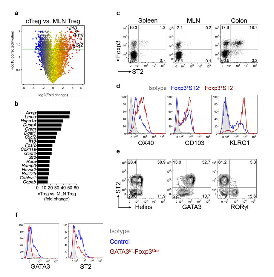
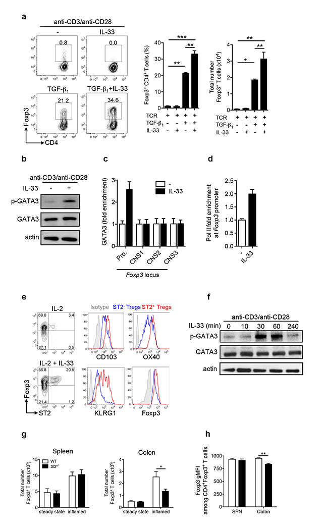
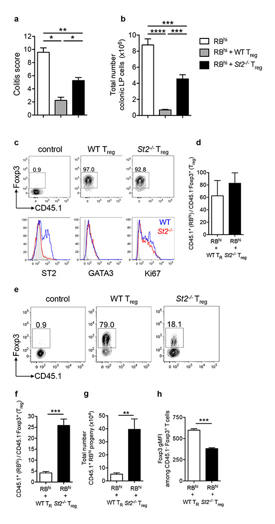
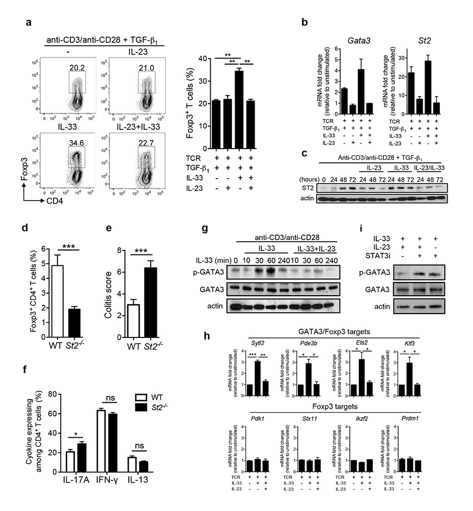
FOXP3(+) regulatory T cells (Treg cells) are abundant in the intestine, where they prevent dysregulated inflammatory responses to self and environmental stimuli. It is now appreciated that Treg cells acquire tissue-specific adaptations that facilitate their survival and function; however, key host factors controlling the Treg response in the intestine are poorly understood. The interleukin (IL)-1 family member IL-33 is constitutively expressed in epithelial cells at barrier sites, where it functions as an endogenous danger signal, or alarmin, in response to tissue damage. Recent studies in humans have described high levels of IL-33 in inflamed lesions of inflammatory bowel disease patients, suggesting a role for this cytokine in disease pathogenesis. In the intestine, both protective and pathological roles for IL-33 have been described in murine models of acute colitis, but its contribution to chronic inflammation remains ill defined. Here we show in mice that the IL-33 receptor ST2 is preferentially expressed on colonic Treg cells, where it promotes Treg function and adaptation to the inflammatory environment. IL-33 signalling in T cells stimulates Treg responses in several ways. First, it enhances transforming growth factor (TGF)-β1-mediated differentiation of Treg cells and, second, it provides a necessary signal for Treg-cell accumulation and maintenance in inflamed tissues. Strikingly, IL-23, a key pro-inflammatory cytokine in the pathogenesis of inflammatory bowel disease, restrained Treg responses through inhibition of IL-33 responsiveness. These results demonstrate a hitherto unrecognized link between an endogenous mediator of tissue damage and a major anti-inflammatory pathway, and suggest that the balance between IL-33 and IL-23 may be a key controller of intestinal immune responses.


Figures

Figure 1
Figure 1. ST2-expressing Treg cells are enriched in the colon
a, Change in gene expression in cTreg cells vs. MLN Treg cells (n=3 per group) presented as volcano plot. b, Top differentially upregulated transcripts in cTreg vs. MLN Treg cells. c, ST2 protein expression on Treg cells from indicated organs. d, Phenotypic analysis of ST2 or ST2+ cTreg cells. e, Expression of transcription factors in cTreg cells. f, Representative histograms gated on cTreg cells from control or Gata3f/f-Foxp3-Cre mice.
Figure 2
Figure 2. Effects of IL-33 on iTreg and tTreg cells
a, Naïve CD4+ T cells were cultured with anti-CD3/CD28 plus indicated cytokines and the frequencies and absolute numbers of Foxp3+ T cells determined 3 days later (mean ± s.e.m. of three independent experiments). b, Naïve CD4+ T cells were cultured for 48h with anti-CD3/CD28 plus TGF-β1 followed by stimulation with IL-33 for 45 minutes. Blots are representative of two independent experiments. c-d, Cells were cultured and stimulated as in (b) and recruitment of GATA3 or RNA polymerase II (Pol II) to the indicated regions assessed by ChIP-qPCR. Data are from one experiment representative of two (mean ± s.d.). e, Representative plots of Treg cells cultured with anti-CD3/CD28 plus indicated cytokines and analyzed after 3 days. Data are representative of three independent experiments. f, Treg cells were cultured with anti-CD3/CD28 for 24h followed by stimulation with IL-33. Blots are representative of three independent experiments. g, Mixed chimaeras were generated containing WT and St2−/− bone marrow cells. Reconstituted mice were analyzed at steady state or two weeks after infection with Helicobacter hepaticus and anti-IL-10R treatment (inflamed). Absolute numbers of WT or St2−/− Treg cells in steady state (n = 3) and inflamed (n = 6) hosts (mean ± s.e.m). h, Analysis of Foxp3 expression in Treg cells from inflamed chimaeric hosts presented as geometric mean fluorescence intensity (gMFI). *P<0.05, **P<0.01 *** P<0.001 as calculated by 1way-ANOVA with Bonferroni post-test or paired Students’ t-test.
Figure 3
Figure 3. IL-33 promotes Treg cell stability and function in vivo
a, C57BL/6 Rag1−/− mice were injected with CD45.1+ naïve T cells alone (RBhi, n = 4) or in combination with WT (n = 4) or St2−/− (n = 6) CD45.1 Treg cells. Mice were sacrificed after 6-8 weeks post transfer and colitis scores are shown. b, Absolute numbers of colon lamina propria (LP) cells from mice in (a). c, C57BL/6 Rag1−/− mice were injected as in (a) and sacrificed at 2 weeks post injection. Representative plots are gated on colonic Treg cell progeny (CD45.1). d, Ratio of RBhi T cell progeny (CD45.1+) to WT or St2−/− Foxp3+ Treg cell progeny (CD45.1) in the colon (n = 5 per group) from mice in (c), (mean ± s.e.m). e, C57BL/6 Rag1−/− mice were injected as in (a) and sacrificed at 8 weeks post injection. Representative plots are gated on colonic Treg cell progeny (CD45.1). f, Ratio of RBhi T cell progeny (CD45.1+) to WT or St2−/− Treg cell progeny (CD45.1) in the colon from mice in (e), (mean ± s.e.m). g, Absolute numbers of RBhi T cell progeny (CD45.1+) in the colon from mice in (e), (mean ± s.e.m). h, Analysis of Foxp3 expression in colonic Foxp3+CD45.1 Treg cells presented as gMFI (mean ± s.e.m). Results are representative of two independent experiments. *P<0.05 **P<0.01 *** P<0.001 **** P<0.0001 as calculated by 1way-ANOVA with Bonferroni post-test or Students’ t-test.
Figure 4
Figure 4. IL-23 inhibits the effects of IL-33 on Treg cells
a, Naïve CD4+ T cells were cultured with anti-CD3/CD28 plus TGF-β1 as well as indicated cytokines and the frequencies of Foxp3+ T cells determined after 3 days later (mean ± s.e.m. of three independent experiments). b, Naïve CD4+ T cells were cultured with anti-CD3/CD28 plus indicated cytokines for 48h. Data are from one experiment representative of two (mean ± s.d.). c, Naïve CD4+ T cells were cultured as indicated. Representative blots of two independent experiments are shown. d, C57BL/6 Il23a−/−Rag1−/− mice were injected with CD45RBhi WT (n = 9) or St2−/− (n = 10) T cells. Mice were sacrificed after 6-8 weeks post transfer and frequencies of Foxp3+CD4+ T cells in colon are shown. e, Colitis scores for mice in (d). f, Expression of indicated cytokines by colonic CD4+ T cells from mice in (d). g, Treg cells were cultured with anti-CD3/CD28 for 24h followed by stimulation with IL-33 in the presence or absence of IL-23. Representative blots of two independent experiments are shown. h, Treg cells were cultured in the presence of anti-CD3/CD28 for 24h and mRNA expression of indicated genes measured following stimulation with IL-33 for 45 minutes in the presence or absence of IL-23 (mean ± s.e.m. of three independent experiments). i, Treg cells were cultured with anti-CD3/CD28 for 24h and representative blots of two independent experiments are shown. *P<0.05, **P<0.01 *** P<0.001 as calculated by 1way-ANOVA with Bonferroni post-test or Students’ t-test.

Comment in

References

    1. Burzyn D, Benoist C, Mathis D. Regulatory T cells in nonlymphoid tissues. Nature immunology. 2013;14:1007–1013. - PMC - PubMed
    1. Pichery M, et al. Endogenous IL-33 is highly expressed in mouse epithelial barrier tissues, lymphoid organs, brain, embryos, and inflamed tissues: in situ analysis using a novel Il-33-LacZ gene trap reporter strain. Journal of immunology. 2012;188:3488–3495. - PubMed
    1. Palmer G, Gabay C. Interleukin-33 biology with potential insights into human diseases. Nature reviews. Rheumatology. 2011;7:321–329. - PubMed
    1. Beltran CJ, et al. Characterization of the novel ST2/IL-33 system in patients with inflammatory bowel disease. Inflammatory bowel diseases. 2010;16:1097–1107. - PubMed
    1. Kobori A, et al. Interleukin-33 expression is specifically enhanced in inflamed mucosa of ulcerative colitis. J Gastroenterol. 2010;45:999–1007. - PubMed

Publication types

MeSH terms

Associated data