. 2014 Sep;76(3):393-402.
doi: 10.1002/ana.24220. Epub 2014 Jul 22.

Carboxyfullerene neuroprotection postinjury in Parkinsonian nonhuman primates

Affiliations

Carboxyfullerene neuroprotection postinjury in Parkinsonian nonhuman primates

Laura L Dugan et al. Ann Neurol. 2014 Sep.

Abstract

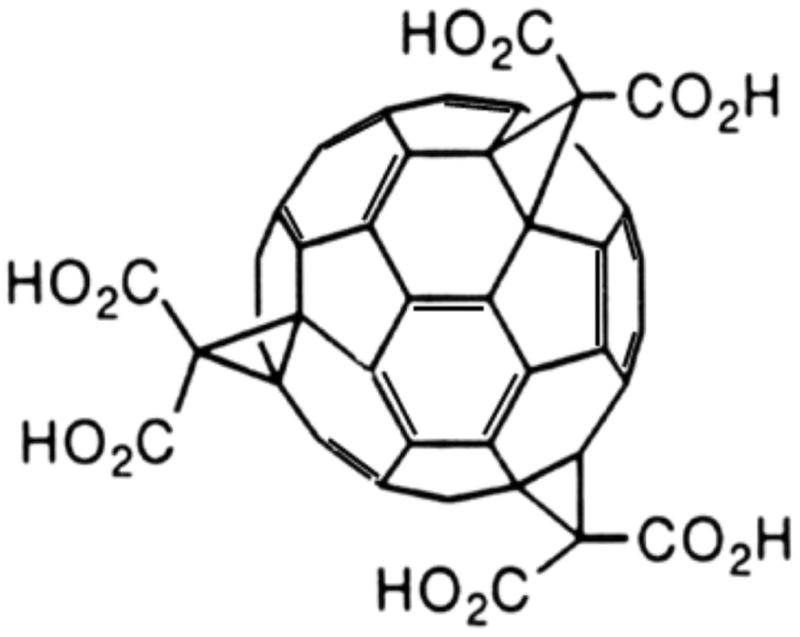
Objective: We evaluated the efficacy of the potent antioxidant C3 to salvage nigrostriatal neuronal function after 1-methyl-4-phenyl-1,2,3,6-tetrahydropyridine (MPTP) exposure in nonhuman primates. C3 is a first-in-class functionalized water-soluble fullerene that reduces oxygen radical species associated with neurodegeneration in in vitro studies. However, C3 has not been evaluated as a neuroprotective agent in a Parkinson model in vivo.

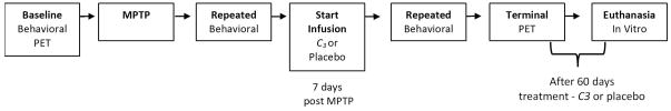
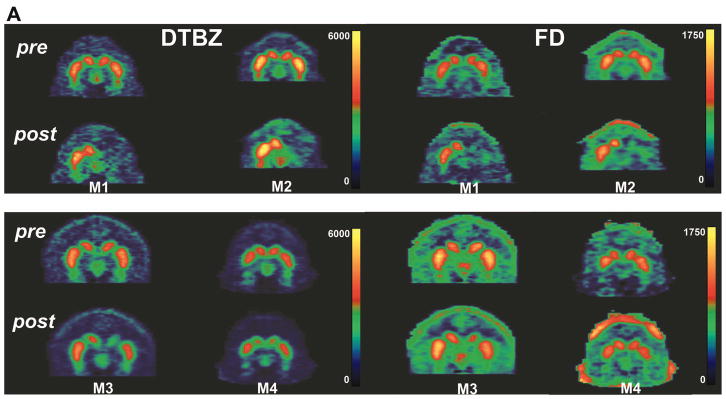
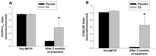
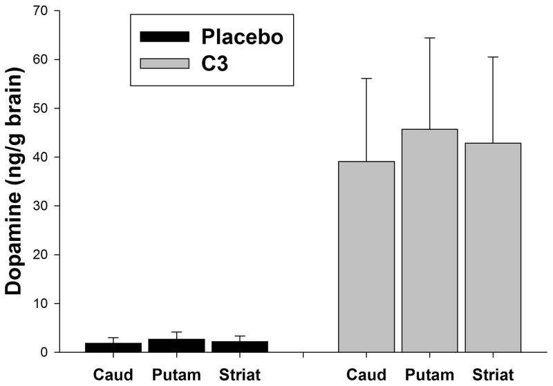
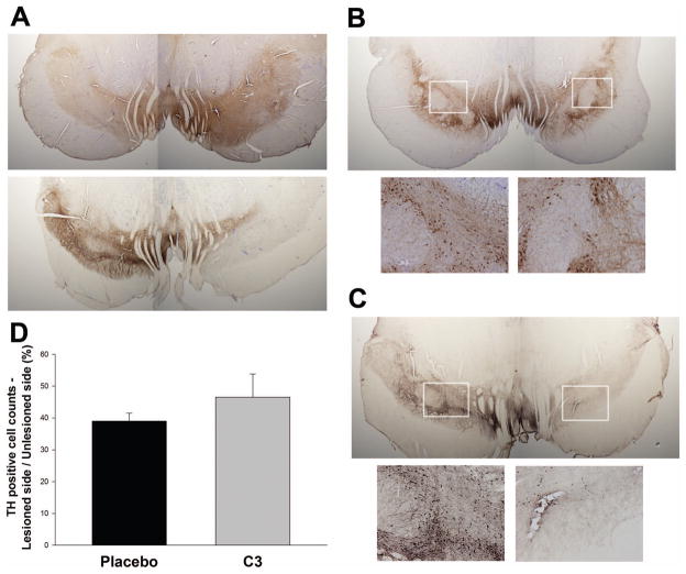
Methods: Macaque fascicularis monkeys were used in a double-blind, placebo-controlled study design. MPTP-lesioned primates were given systemic C3 (n = 8) or placebo (n = 7) for 2 months starting 1 week after MPTP. Outcomes included in vivo behavioral measures of motor parkinsonism using a validated nonhuman primate rating scale, kinematic analyses of peak upper extremity velocity, positron emission tomography imaging of 6-[(18) F]fluorodopa (FD; reflects dopa decarboxylase) and [(11) C]dihydrotetrabenazine (DTBZ; reflects vesicular monoamine transporter type 2), ex vivo quantification of striatal dopamine, and stereologic counts of tyrosine hydroxylase-immunostained neurons in substantia nigra.

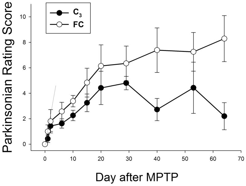
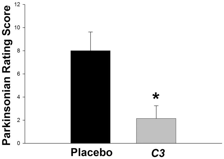
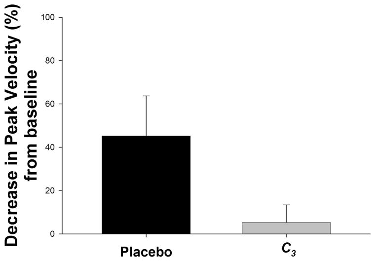
Results: After 2 months, C3 -treated monkeys had significantly improved parkinsonian motor ratings, greater striatal FD and DTBZ uptake, and higher striatal dopamine levels. None of the C3 -treated animals developed any toxicity.

Interpretation: Systemic treatment with C3 reduced striatal injury and improved motor function despite administration after the MPTP injury process had begun. These data strongly support further development of C3 as a promising therapeutic agent for Parkinson disease.


Figures

Figure 1
Figure 1. Chemical structure of C3
Molecular structure showing the three malonic acid groups of C3 attached to the fullerene C60 sphere through cyclopropane carbons at the e, e, e, positions of C60, giving the molecule C3 symmetry. The C3 symmetry gives the compound dipolar character, which enhances distribution across neuronal cell membranes.
Figure 2
Figure 2. Schematic of Study Design
Illustration of the major aspects of the study. Baseline measures (behavioral, PET) precede treatment with intracarotid MPTP. One week after MPTP administration, each animal is assessed to verify nigrostriatal damage, and randomized to either a treatment (C3) or placebo infusion for 8 weeks. Repeated behavioral measures are made during the 8 weeks of infusion. At the end of the infusion interval, terminal PET measures are done, the animal euthanized and in vitro measures (DA, TH) are performed. Note that the process for the treatment and control arms are identical, and that both infusions begin well after (7 days) administration of neurotoxin.
Figure 3
Figure 3. Dynamic functional assessment of Parkinson symptoms for C3-treated and control animals
Animals were rated using an 18-point system which assesses the severity of parkinsonian symptoms (0= no symptoms; 18= most severely affected). Data are the mean and standard deviation of scores for all animals in each arm. Note the similarity of scores shortly after MPTP treatment, but prior to starting infusion of C3 or placebo, and the progressively different scores attained with longer infusions of C3. Scores for the two groups of animals diverged significantly beginning 30 days after the start of C3 infusion (p=0.026 by Mann-Whitney-Wilcoxon). Filled circles, C3-treated animals; open circles, control animals.
Figure 4
Figure 4. Parkinsonian motor scores in the primates at the end of the study
Data represent the mean ± SEM at the end of the 8-week infusions. At the end of 2 months of treatment, C3-treated monkeys had a significantly lower rating compared to controls (Mann-Whitney-Wilcoxon rank sum test: U=4.5, n1 = n2 = 7, p = 0.007).
Figure 5
Figure 5. Kinematic scores of upper extremity peak velocity
Kinematic data are analyzed as the ratio of the peak movement velocity for the affected (right) upper extremity in animals at the end of the 8-week treatment compared to that of the same limb prior to MPTP lesioning. Results are presented as the mean percent decrease in velocity from baseline, with a smaller decrease indicating preservation of function. Data represent the means ± SEM at the end of the 8-week infusions; n = 8 for C3 and n = 7 for placebo. Compared to controls, C3-treated monkeys appeared to retain upper extremity movement speed (less bradykinesia), but this did not quite reach statistical significance (p = 0.078) by ANOVA t test).
Figure 6
Figure 6. PET imaging of DTBZ and FD
Representative coronal PET images are shown from two control primates (A; monkeys M1 and M2) and two C3-treated monkeys (B; monkeys M3 and M4) just prior to intracarotid MPTP (top panels) and at the end of two months of treatment with placebo or C3 (bottom panels). Images of DTBZ (left panels) and FD (right panels) are shown. Note the bilateral uptake of each tracer pre-MPTP, showing the tear-drop-shaped substantia nigra bilaterally for all four animals. However, at the end of the study, there is significantly less uptake of both tracers on the lesioned side in placebo-treated animals compared to C3-treated monkeys, or conversely, there is preservation of DTBZ and FD in C3-treated animals.
Figure 7
Figure 7. Summary of PET measures after completion of C3 or placebo infusion
Quantification of (A) FD Kocc and (B) DTBZ BP at the end of the study. Data are the mean ± SEM of the nigral tracer uptake before MPTP, and at the end of the 2-month drug or placebo infusion. There was significant preservation of uptake for both tracers (FP Kocc p=0.038; DTBZ p=0.02) in the C3-treated group by repeated measures ANOVA, with Tukey’s post-hoc test. C3, n = 8; controls, n = 7.
Figure 8
Figure 8. Striatal dopamine levels
Tissue dopamine concentration per mg wet weight tissue was determined by HPLC (a) at the completion of the study, following the 2-month drug treatment. The concentration of dopamine in the lesioned caudate (caud), putamen (put), and striatum (striat) was divided by the concentration in the non-lesioned side to control for differences in brain size or time to dissect the tissue. There was significant (p < 0.001) preservation of dopamine in all three regions in C3-treated animals compared to placebo-treated controls (2-way ANOVA, Tukey’s post-hoc test).
Figure 9
Figure 9. Imaging of tyrosine hydroxylase (TH) immunoreactive neurons and processes in substantia nigra in placebo and C3 treated monkeys
Brains were sliced and immunostained for TH using DAB amplification. TH is shown in brown, with some slices counterstained with cresyl violet. The corresponding left and right (MPTP lesioned) hemispheres from each animal are shown from two independent pairs of monkeys. (A) Comparison of a placebo and a C3 treated monkey showing substantial loss of TH immunoreactivity on the lesioned side with placebo, and less loss in the C3-treated animal. (B, C) different pair of monkeys showing similar loss of TH positive neurons (magnified in insets) in the C3 treated animal, which is more dramatic in the placebo-treated animals. (D) Quantification showing a nonsignificant trend (p = 0.078, by ANOVA) toward preservation of nigral TH cell bodies by stereological counts. Qualitatively, the degree of preservation of TH immunoreactivity in neuronal processes appears substantially greater than that in cell bodies.

References

    1. Ali SS, Hardt JI, Quick KL, Kim-Han JS, Erlanger BF, Huang TT, Epstein CJ, Dugan LL. A biologically effective fullerene (C60) derivative with superoxide dismutase mimetic properties. Free Radic Biol Med. 2004;37:1191–1202. - PubMed
    1. Anderson DG, Mariappan SV, Buettner GR, Doorn JA. Oxidation of 3,4-dihydroxyphenylacetaldehyde, a toxic dopaminergic metabolite, to a semiquinone radical and an ortho-quinone. J Biol Chem. 2011;286:26978–26986. - PMC - PubMed
    1. Behrens MM, Ali SS, Dugan LL. Interleukin-6 mediates the increase in NADPH-oxidase in the ketamine model of schizophrenia. J Neurosci. 2008;28:13957–13966. - PMC - PubMed
    1. Behrens MM, Ali SS, Dao DN, Lucero J, Shekhtman G, Quick KL, Dugan LL. Ketamine-induced loss of phenotype of fast-spiking interneurons Is mediated by NADPH-oxidase. Science. 2007;318:1645–1647. - PubMed
    1. Bezard E, Przedborski S. A tale on animal models of Parkinson’s disease. MovDisord. 2011;26:993–1002. - PubMed

Publication types

MeSH terms