Case Reports
. 2014 Oct;35(10):1153-62.
doi: 10.1002/humu.22618. Epub 2014 Aug 11.

Mutation of POC1B in a severe syndromic retinal ciliopathy

Affiliations
Case Reports

Mutation of POC1B in a severe syndromic retinal ciliopathy

Bodo B Beck et al. Hum Mutat. 2014 Oct.

Abstract

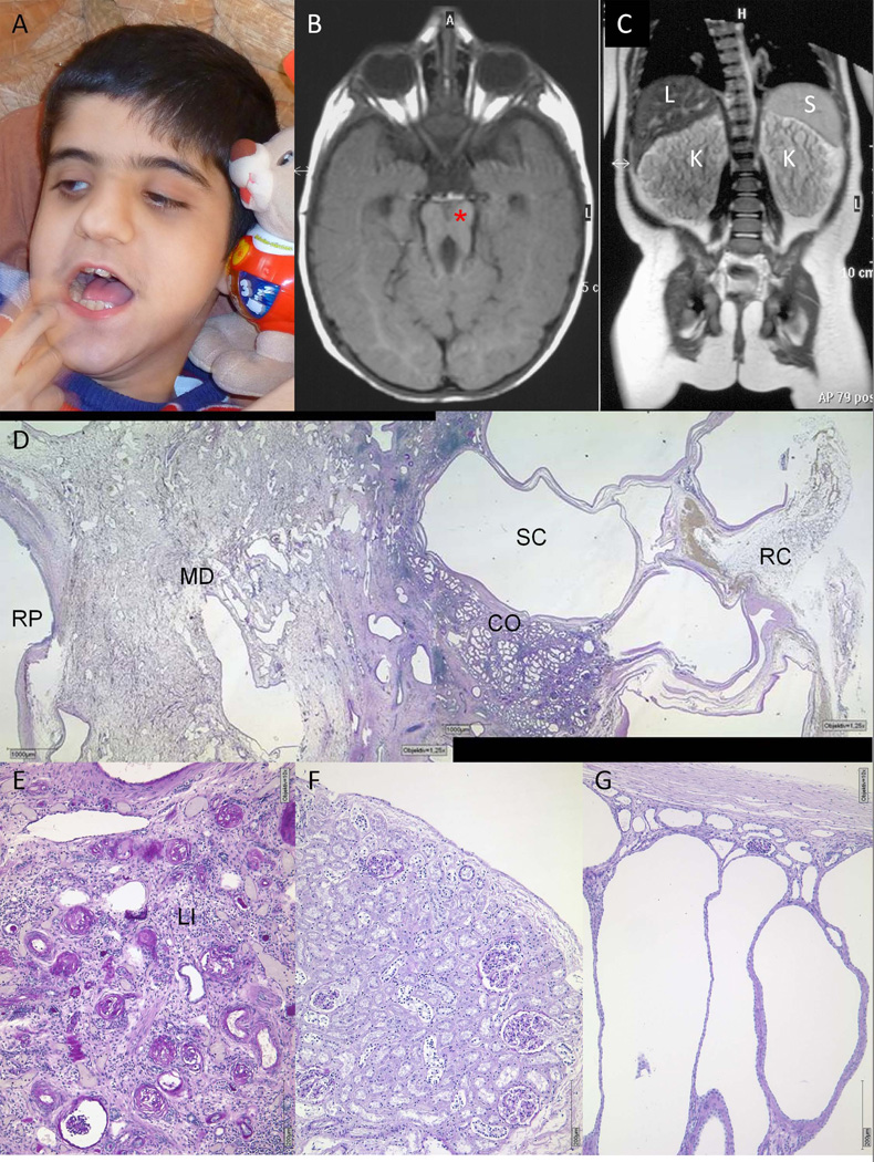
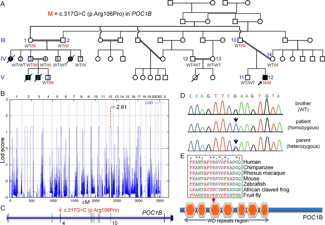
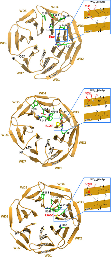
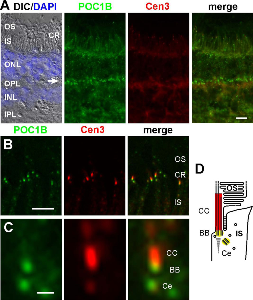
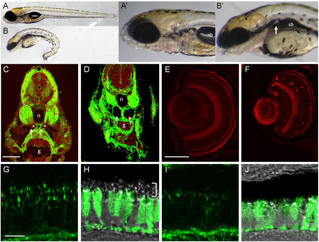
We describe a consanguineous Iraqi family with Leber congenital amaurosis (LCA), Joubert syndrome (JBTS), and polycystic kidney disease (PKD). Targeted next-generation sequencing for excluding mutations in known LCA and JBTS genes, homozygosity mapping, and whole-exome sequencing identified a homozygous missense variant, c.317G>C (p.Arg106Pro), in POC1B, a gene essential for ciliogenesis, basal body, and centrosome integrity. In silico modeling suggested a requirement of p.Arg106 for the formation of the third WD40 repeat and a protein interaction interface. In human and mouse retina, POC1B localized to the basal body and centriole adjacent to the connecting cilium of photoreceptors and in synapses of the outer plexiform layer. Knockdown of Poc1b in zebrafish caused cystic kidneys and retinal degeneration with shortened and reduced photoreceptor connecting cilia, compatible with the human syndromic ciliopathy. A recent study describes homozygosity for p.Arg106ProPOC1B in a family with nonsyndromic cone-rod dystrophy. The phenotype associated with homozygous p.Arg106ProPOC1B may thus be highly variable, analogous to homozygous p.Leu710Ser in WDR19 causing either isolated retinitis pigmentosa or Jeune syndrome. Our study indicates that POC1B is required for retinal integrity, and we propose POC1B mutations as a probable cause for JBTS with severe PKD.

Keywords: Joubert syndrome; LCA; POC1B; ciliopathy; zebrafish.


Conflict of interest statement

CONFLICT OF INTEREST STATEMENT

T.E., C.B. and H.J.B. are employees of Bioscientia which is part of a publicly traded diagnostic company. The other authors have no competing interests.

Figures

FIGURE 1
FIGURE 1. Clinical findings in the index patient
A Facial appearance without gross dysmorphology. B Axial brain MRI showing molar tooth sign (*). C MRI of the trunk showing massively enlarged polycystic kidneys with loss of corticomedullary differentiation. K, kidney; L, liver; S, spleen. D, E Specimen from right nephrectomy (269 g; 10,5 × 6,5 × 7,5 cm) at the age of 4.5 years (PAS; D, 12.5×; E, 100×). D Overview showing completely abolished tissue architecture with tall (Ø up to 1,2 cm) fluid-filled subcapsular cysts (SC) of tubular origin mainly in cortical (CO) and subcapsular localization. The renal pelvis (RP) was dilated and covered by normal urothelial layer. MD, medulla; RC, renal capsule. E High magnification of cortical region with obsolescent glomeruli, lymphohistioplasmocytic infiltration (LI), severe tubular atrophy and interstitial fibrosis. Hypertrophic tubular structures resemble distal tubuli. Note almost complete absence of collecting ducts and loop of Henle structures in the medullary area (MD). Glomeruli were either normal or compatible with unspecific obsolescence, and arterioles and interlobular arteries appeared normal (not shown). F Regular renal cortex (PAS, 100×) for comparison. G For comparison, a typical histology of an ARPKD patient (PAS, 100×) is shown with elongated cysts reaching from the cortex to the medulla, separated by loose connective tissue with interspersed glomerular structures.
FIGURE 2
FIGURE 2. Genetic characterization of the consanguineous Iraqi family segregating a POC1B mutation
A Pedigree. The sample of the index patient (arrow) was submitted to exome sequencing. For detailed clinical data of the different individuals, see Supp. Table S2. POC1B genotypes: WT, wild-type; M, mutant (c.317G>C). B Graphical view of the LOD score calculation of genome-wide SNP mapping (based on samples from III:10, IV:14, V:11 and V:12). Regions showing HBD were identified on chromosomes 2 (2×), 6, 7, 9, 10, 12, 13, 15, 17 (2×) and 19. The dotted line indicates the LOD score for c.317G>C in POC1B based on genotypes from all finally available samples in the family. C Scheme of the POC1B gene with the identified mutation. D Sanger sequencing confirmed the homozygous mutation, c.317G>C (p.Arg106Pro) in POC1B exon 4, in the patient (middle panel). It was found in heterozygous state in both parents (lower panel), while the healthy brother displayed wild-type sequence (upper panel). Scheme of the POC1B protein with the mutation affecting a evolutionarily highly conserved residue in the third WD40 repeat. For clarity, the one-letter amino acid code was applied for mutation designation in this subset of the figure.
FIGURE 3
FIGURE 3. Structure of wild-type, mutant and variant POC1B WD40 domain as predicted by the WDSP algorithm
(refined by an 800 ns MD simulation using a residue specific force-field). For simplicity, the one-letter-code is being used for amino acid residues. The strands a–d are depicted in WD1, and the WDb-a β-bulge is shown for WD3. A The seven WD40 blades are well defined in the wild-type, each exhibiting a β-bulge formed between the beginning of strand a and the end of strand b (WDb-a bulge). The side chain of p.Arg106, which locates at the R1 position of the bulge in the blade WD3, extrudes on the top face of the propeller. p.Arg106 and several other residues shown in the figure are predicted to be the potential hotspots for ligand binding. B The p.Arg106Pro mutation disrupts the WDb-a bulge in WD3, perturbs the conformation of p.Trp146, p.Arg80 and p.Glu122 and the ligand binding environment of the top face of the propeller. C The known p.Arg106Gln polymorphism (SNP rs76216585) only has a minor effect on structure. The side chains of Arg and Gln are both extended. A hydrogen bond between p.Gln106 and p.Glu122 in the variant protein ould provide electrostatic stabilization, similar the salt bridge between p.Arg106 and p.Glu122 in the wild-type protein.
FIGURE 4
FIGURE 4. Localization of POC1B in the human retina
A Longitudinal cryosections through a human retina stained for POC1B (green) and counterstained for the ciliary marker centrin-3 (Cen3, red) reveals POC1B localization in the ciliary region (CR) of the photoreceptor layer and the synapses of the outer plexiform layer (OPL) (arrow). Overlay of DIC (differential interference contrast) image with DAPI (blue) nuclear stain in the outer (ONL) and inner nuclear layer (INL) shows retina layers. OS, outer segment; IS, inner segment. B Increased magnification of the photoreceptor cells demonstrates co-localization of POC1B and Cen3 in the photoreceptor ciliary region (CR). C High magnification of the photoreceptor cilium reveals substantial localization of POC1B at the centriole (Ce) and the basal body (BB) of the connecting cilium (CC), but not in the CC itself, as schematically demonstrated in D. Scale bars: A, 10 µm; B, 5 µm; C, 0.5 µm.
FIGURE 5
FIGURE 5. Morpholino knockdown of zebrafish poc1b results in photoreceptor abnormalities and retinal cell death
A – B whole 5dpf larvae, lateral views. A’, B’ High magnification views of the animals pictured in A and B. Compared to controls (A), morpholino treated larvae (B) have curved body axes, small eyes and heads and reduced body length. The swim bladder (sb) does not inflate in poc1b morphants and kidney cysts are observed in a subset of poc1bMO animals (white arrow in B’). C – D Transverse sections through the trunk region of 4dpf control (C) and morphant (D) larvae labeled with actin (green) and TOPRO3 (red). Pronephric ducts are indicated with asterisks in C; white arrowheads indicate kidney cysts in the poc1bMO animal. E – F Retinal cell death in poc1bMO larvae. Sectioned retinal tissue from 5dpf control animals (E) labeled with Caspase-3 (red) are negative for retinal cell death at this stage. Some autofluorescence is present in the photoreceptor outer segments. poc1bMO retinal sections (F) show normal retinal cell layers but smaller eye size relative to controls and an increase in retinal cell death, primarily in the inner nuclear layer. G – J Photoreceptor connecting cilia and outer segments are abnormal in poc1b morphants. Compressed 10µm z-stack of retinas stained with acetylated tubulin (G, I) show the tiered arrangement and length of connecting cilia in control retinas (G). Cilia are reduced in length and number in the morphant retinas (I). Green signal at the base of the images marks the outer plexiform layer. H, J The zpr1 antibody (green) labels the inner segments of the long red/green double cones. Uniform distribution of zpr1 from pedicles to the top of the inner segment is observed in controls (H). DIC imaging shows cone outer segments (white bracket) superior to the zpr1 label and inferior to the retinal pigmented epithelium. In poc1b morphants (J), inner segments appear somewhat reduced by zpr1 labeling, and few outer segments are observed by DIC. Scale bars: C–F: 50 µm; G–J: 10 µm. e, ear; sb, swim bladder; n, notochord; g, gut.

References

    1. Basmanav FB, Oprisoreanu AM, Pasternack SM, Thiele H, Fritz G, Wenzel J, Grosser L, Wehner M, Wolf S, Fagerberg C, Bygum A, Altmuller J, et al. Mutations in POGLUT1, encoding protein O-glucosyltransferase 1, cause autosomal-dominant Dowling-Degos disease. Am J Hum Genet. 2014;94:135–143. - PMC - PubMed
    1. Boldt K, Mans DA, Won J, van Reeuwijk J, Vogt A, Kinkl N, Letteboer SJ, Hicks WL, Hurd RE, Naggert JK, Texier Y, den Hollander AI, et al. Disruption of intraflagellar protein transport in photoreceptor cilia causes Leber congenital amaurosis in humans and mice. J Clin Invest. 2011;121:2169–2180. - PMC - PubMed
    1. Bredrup C, Saunier S, Oud MM, Fiskerstrand T, Hoischen A, Brackman D, Leh SM, Midtbo M, Filhol E, Bole-Feysot C, Nitschke P, Gilissen C, et al. Ciliopathies with skeletal anomalies and renal insufficiency due to mutations in the IFT-A gene WDR19. Am J Hum Genet. 2011;89:634–643. - PMC - PubMed
    1. Coussa RG, Otto EA, Gee HY, Arthurs P, Ren H, Lopez I, Keser V, Fu Q, Faingold R, Khan A, Schwartzentruber J, Majewski J, et al. WDR19: an ancient, retrograde, intraflagellar ciliary protein is mutated in autosomal recessive retinitis pigmentosa and in Senior-Loken syndrome. Clin Genet. 2013;84:150–159. - PMC - PubMed
    1. Durlu YK, Koroglu C, Tolun A. Novel Recessive Cone-Rod Dystrophy Caused by POC1B Mutation. JAMA Ophthalmol. 2014 - PubMed

Publication types

MeSH terms

Supplementary concepts