

Planum temporale asymmetry in developmental dyslexia: Revisiting an old question
- PMID: 25044828
- PMCID: PMC6869664
- DOI: 10.1002/hbm.22579
Planum temporale asymmetry in developmental dyslexia: Revisiting an old question
Abstract
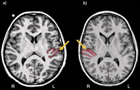
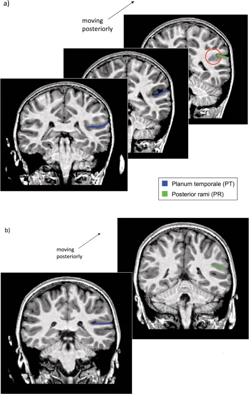
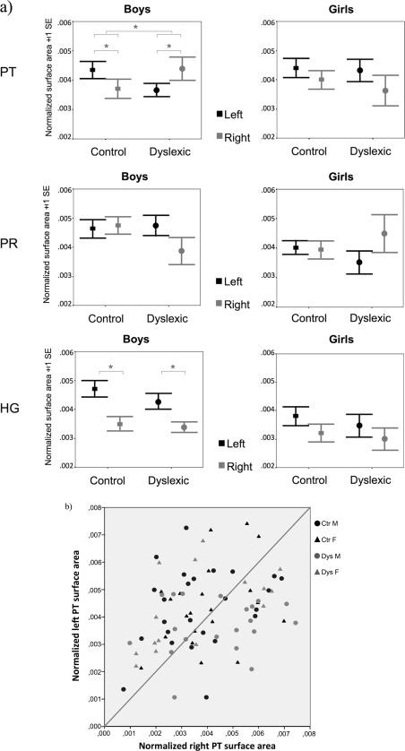
Among the various asymmetrical structures of the human brain, the planum temporale, an anatomical region associated with a variety of auditory and language-related processes, has received particular attention. While its surface area has been shown to be greater in the left hemisphere compared to the right in about two-thirds of the general population, altered patterns of asymmetry were revealed by post mortem analyses in individuals with developmental dyslexia. These findings have been inconsistently replicated in magnetic resonance imaging studies of this disorder. In this report, we attempt to resolve past inconsistencies by analyzing the T1-weighted MR images of 81 children (mean age: 11 years, sd: 17 months), including 46 control (25 boys) and 35 dyslexic children (20 boys). We manually outlined Heschl's gyri, the planum temporale and the posterior rami of the Sylvian fissure on participants' brain images, using the same anatomical criteria as in post mortem studies. Results revealed an altered pattern of asymmetry of the planum temporale surface area in dyslexic boys only, with a greater proportion of rightward asymmetrical cases among dyslexic boys compared to control boys. Additionally, analyses of cortical thickness showed no asymmetry differences between groups for any of the regions of interest. Finally, a greater number of Heschl's gyrus full duplications emerged for the right hemisphere of dyslexic boys compared to controls. The present findings confirm and extend early post mortem observations. They also stress the importance of taking gender into account in studies of developmental dyslexia.
Keywords: developmental dyslexia; gender; planum temporale; reading; structural magnetic resonance imaging.
© 2014 Wiley Periodicals, Inc.
Figures


References
-
- Barta PE, Pearlson GD, Brill LB 2nd, Royall R, McGilchrist IK, Pulver AE, Powers RE, Casanova MF, Tien AY, Frangou S, Petty RG (1997): Planum temporale asymmetry reversal in schizophrenia: Replication and relationship to gray matter abnormalities. Am J Psychiatry 154:661–667. - PubMed
-
- Best M, Demb JB (1999): Normal planum temporale asymmetry in dyslexics with a magnocellular pathway deficit. Neuroreport 10:607–612. - PubMed
-
- Buxhoeveden D, Switala A, Litaker M, Roy E, Casanova MF (2001): Lateralization of minicolumns in human planum temporale is absent in nonhuman primate cortex. Brain Behav Evol 57:349–358. - PubMed
-
- Casanova MF, Buxhoeveden DP, Cohen M, Switala AE, Roy EL (2002): Minicolumnar pathology in dyslexia. Ann Neurol 52:108–110. - PubMed
Publication types
MeSH terms
LinkOut - more resources
Full Text Sources
Other Literature Sources
Miscellaneous
