Review
. 2014 Jul 5:7:183-97.
doi: 10.2147/IDR.S39601. eCollection 2014.

Actinomycosis: etiology, clinical features, diagnosis, treatment, and management

Affiliations
Review

Actinomycosis: etiology, clinical features, diagnosis, treatment, and management

Florent Valour et al. Infect Drug Resist. .

Abstract

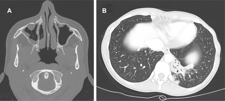
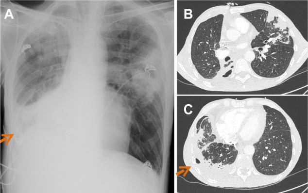
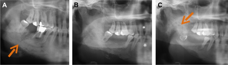
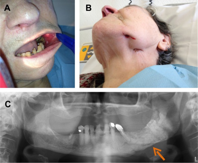
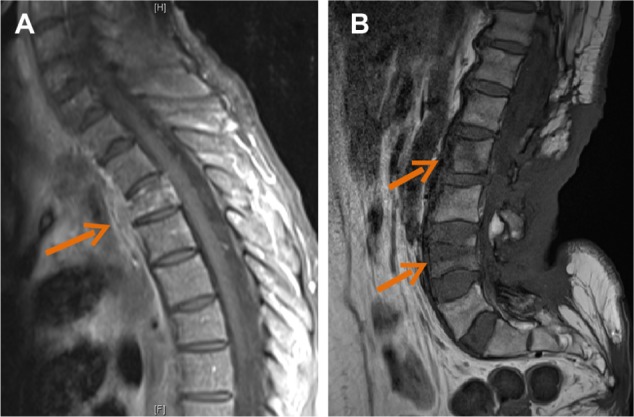
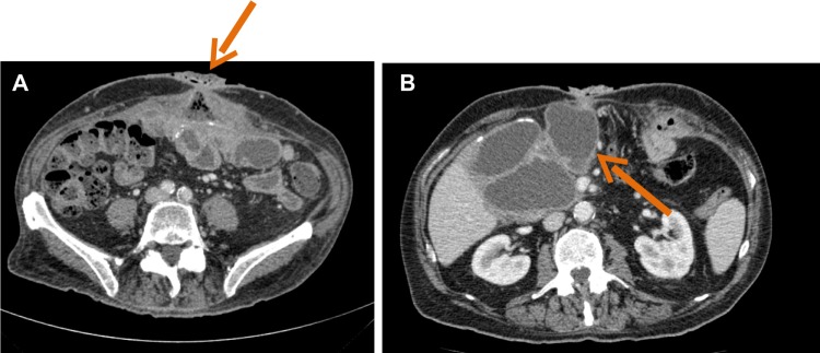
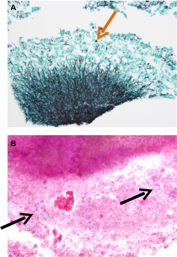
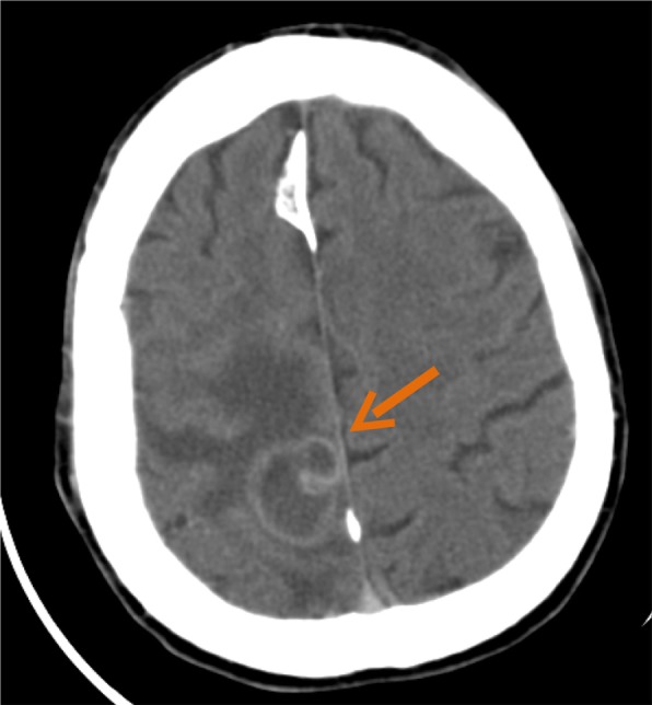
Actinomycosis is a rare chronic disease caused by Actinomyces spp., anaerobic Gram-positive bacteria that normally colonize the human mouth and digestive and genital tracts. Physicians must be aware of typical clinical presentations (such as cervicofacial actinomycosis following dental focus of infection, pelvic actinomycosis in women with an intrauterine device, and pulmonary actinomycosis in smokers with poor dental hygiene), but also that actinomycosis may mimic the malignancy process in various anatomical sites. Bacterial cultures and pathology are the cornerstone of diagnosis, but particular conditions are required in order to get the correct diagnosis. Prolonged bacterial cultures in anaerobic conditions are necessary to identify the bacterium and typical microscopic findings include necrosis with yellowish sulfur granules and filamentous Gram-positive fungal-like pathogens. Patients with actinomycosis require prolonged (6- to 12-month) high doses (to facilitate the drug penetration in abscess and in infected tissues) of penicillin G or amoxicillin, but the duration of antimicrobial therapy could probably be shortened to 3 months in patients in whom optimal surgical resection of infected tissues has been performed. Preventive measures, such as reduction of alcohol abuse and improvement of dental hygiene, may limit occurrence of pulmonary, cervicofacial, and central nervous system actinomycosis. In women, intrauterine devices must be changed every 5 years in order to limit the occurrence of pelvic actinomycosis.

Keywords: Actinomyces spp.; lumpy jaw syndrome; osteomyelitis; sulfur granule.


Figures

Figure 1
Figure 1
Computed tomography scan revealing a right maxillary sinusitis (A) and left focal basal pneumonia without cavitation (B), due to Actinomyces spp., in an immunosuppressed woman.
Figure 2
Figure 2
Chest X-ray (A) and thorax computed tomography scan (BC) revealing multifocal pneumonia with right pleural cavitation due to Actinomyces viscosus. Note: Arrows indicate chest wall sinus tract.
Figure 3
Figure 3
Panoramic dental X-ray showing right mandibular osteomyelitis (“lumpy jaw syndrome”). Notes: An unfavorable outcome resulted, despite dental extraction of tooth 46 (with arrow showing mandibular thickening with lucencies) (A), followed by tooth 45 2 months later (B). Surgical debridement and decortication were required due to extension of the osteomyelitis to the gonial angle and to the ramus (with arrow showing the typical radiologic aspect of lumpy jaw syndrome) (C).
Figure 4
Figure 4
Left mandibular osteomyelitis with bone exposure (A) and sinus tract (B) following left mandibular radiotherapy in a patient receiving long-term bisphosphonate therapy. Panoramic dental X-ray shows mandibular lucencies (C). Note: The arrow shows mandibular lucencies.
Figure 5
Figure 5
Contrast-enhanced magnetic resonance images showing contiguous spread of pulmonary actinomycosis to the spine (case 2), with thoracic spondylitis of the T3 vertebral body, associated with anterior paravertebral abscess (arrow) (A). Magnetic resonance image showing back soft tissue infiltration, with posterior epiduritis and infection of L2 and L4 vertebral bodies (arrows) in a paraplegic patient with plurimicrobial bone and joint infection following chronic back scar (case 6) (B).
Figure 6
Figure 6
Abdominal computed tomography scan showing peritoneal effusion and heterogeneous pelvic mass surrounding an intrauterine device (A), with abscesses (B) corresponding with pelvic actinomycosis.
Figure 7
Figure 7
Abdominal computed tomography scan of a patient with evidence of actinomycosis on pathology. Notes: Enterocutaneous fistula (arrow) (A) was associated with large intra-abdominal abscess (arrow) (B).
Figure 8
Figure 8
Pathology of a suppurative lesion in a patient with an abdominal wall implant. Notes: Aggregates of Actinomyces spp. and sulfur granules (arrow) after silver staining (A); filamentous bacteria (arrows) after Gram staining (B).
Figure 9
Figure 9
Computed tomography scan revealing a right temporoparietal abscess (arrow) with perilesional edema. Note: Actinomyces meyeri was found in cultures.
Figure 10
Figure 10
Contrast-enhanced magnetic resonance image of the left ear of a patient with an abscess (arrow) due to Actinomyces spp. earlobe infection following acupuncture.

References

    1. Wong VK, Turmezei TD, Weston VC. Actinomycosis. BMJ. 2011;343:d6099. - PubMed
    1. Smego RA, Jr, Foglia G. Actinomycosis. Clin Infect Dis. 1998;26(6):1255–1261. - PubMed
    1. Mandell GL, Bennett JE, Dolin R, editors. Mandell, Douglas, and Bennett’s Principles and Practice of Infectious Diseases. 7th ed. Philadelphia, PA: Churchill Livingstone Elsevier; 2010.
    1. Schaal KP, Lee HJ. Actinomycete infections in humans – a review. Gene. 1992;115(1–2):201–211. - PubMed
    1. Pulverer G, Schütt-Gerowitt H, Schaal KP. Human cervicofacial actinomycoses: microbiological data for 1997 cases. Clin Infect Dis. 2003;37(4):490–497. - PubMed

LinkOut - more resources