. 2014 May;28(2):58-63.
doi: 10.1055/s-0034-1376263.

Vascular malformations: a review

Affiliations

Vascular malformations: a review

Joshua A Cox et al. Semin Plast Surg. 2014 May.

Abstract

Identification and treatment of vascular malformations is a challenging endeavor for physicians, especially given the great concern and anxiety created for patients and their families. The goal of this article is to provide a review of vascular malformations, organized by subtype, including capillary, venous, lymphatic and arteriovenous malformations. Only by developing a clear understanding of the clinical aspects, diagnostic tools, imaging modalities, and options for intervention will appropriate care be provided and results maximized.

Keywords: arteriovenous malformation; capillary malformation; lymphatic malformation; vascular malformation; venous malformation.


Figures

Fig. 1
Fig. 1
Mixed lymphatico-venous malformation (LVM) involving lower extremity. (A) Ulceration is an indication for intervention. (B) Axial, contrast-enhanced magnetic resonance image shows both irregular but extensive, contrast-enhancing lesion indicative of a venous malformation of the lateral thigh as well as (C) cystic, nonenhancing densities along gluteus muscle and pararectal area indicative of a lymphatic malformation.
Fig. 2
Fig. 2
Very large lymphatic malformation involving the right head and neck region and compressing the airway.
Fig. 3
Fig. 3
A 19-year-old man with a capillary malformation in V1 and V2 distribution, status post-serial laser treatments, lip debulking, and excision of redundant tissue through alar and upper lip incision.
Fig. 4
Fig. 4
Various manifestations of localized venous malformations. Venous malformations are bluish, soft, compressible lesions typically found on the (A,B) face, (C) limbs, or trunk.
Fig. 5
Fig. 5
Venous malformation involving a volar forearm. (A,B) Magnetic resonance imaging (coronal and axial views) shows a contrast-enhancing lesion. (C) Clinically, the mass was bluish, soft, and compressible. (D) Pathologic examination confirmed this mass to be a venous malformation.
Fig. 6
Fig. 6
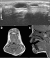
A 4-year-old boy with lymphatic malformations (LMs) involving the chin, tongue, and sublingual spaces. (A) Ultrasound shows avascular, cystic soft tissue masses consistent with an LM. (B) Axial and (C) sagittal views of contrast-enhanced magnetic resonance imaging show a nonenhancing bilobed chin mass as well as a multispacial lesion involving the tongue and sublingual spaces.
Fig. 7
Fig. 7
A 4-year-old boy with lymphatic malformation involving the (A,B) chin, (C,D) status postradical resection.
Fig. 8
Fig. 8
A 12-year-old boy with biopsy-proven right periorbital arteriovenous malformation (AVM) causing (A) vertical dystopia and proptosis. (B) Angiogram was obtained confirming AVM, but was not amenable to embolization due to involvement of the ophthalmic artery. (C) Given the bony involvement, CT scan was useful for operative planning. (D) A radical resection was performed with subsequent orbital roof and forehead reconstruction with alloplast. (E,F) Vertical dystopia and proptosis was improved postoperatively.

References

    1. Mulliken J B, Glowacki J. Classification of pediatric vascular lesions. Plast Reconstr Surg. 1982;70(1):120–121. - PubMed
    1. Van Aalst J A, Bhuller A, Sadove A M. Pediatric vascular lesions. J Craniofac Surg. 2003;14(4):566–583. - PubMed
    1. Mulliken J B, Glowacki J. Hemangiomas and vascular malformations in infants and children: a classification based on endothelial characteristics. Plast Reconstr Surg. 1982;69(3):412–422. - PubMed
    1. Cohen M M Jr. Vascular update: morphogenesis, tumors, malformations, and molecular dimensions. Am J Med Genet A. 2006;140(19):2013–2038. - PubMed
    1. Lowe L H, Marchant T C, Rivard D C, Scherbel A J. Vascular malformations: classification and terminology the radiologist needs to know. Semin Roentgenol. 2012;47(2):106–117. - PubMed