Review
. 2014 Mar 19:2:e28426.
doi: 10.4161/tisb.28426. eCollection 2014.

Structure and function of the ependymal barrier and diseases associated with ependyma disruption

Affiliations
Review

Structure and function of the ependymal barrier and diseases associated with ependyma disruption

Antonio J Jiménez et al. Tissue Barriers. .

Abstract

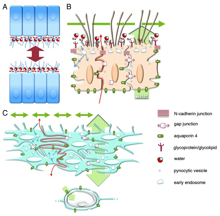
The neuroepithelium is a germinal epithelium containing progenitor cells that produce almost all of the central nervous system cells, including the ependyma. The neuroepithelium and ependyma constitute barriers containing polarized cells covering the embryonic or mature brain ventricles, respectively; therefore, they separate the cerebrospinal fluid that fills cavities from the developing or mature brain parenchyma. As barriers, the neuroepithelium and ependyma play key roles in the central nervous system development processes and physiology. These roles depend on mechanisms related to cell polarity, sensory primary cilia, motile cilia, tight junctions, adherens junctions and gap junctions, machinery for endocytosis and molecule secretion, and water channels. Here, the role of both barriers related to the development of diseases, such as neural tube defects, ciliary dyskinesia, and hydrocephalus, is reviewed.

Keywords: Ependyma; aquaporin 4; astrocyte reaction; cell junctions; cilia; development; hydrocephalus; neural tube defects.


Figures

None
Figure 1. Development and properties of the multiciliated ependyma in the lateral ventricle of human fetuses. (A, insert) In a fetus at 23 wk of gestation, radial glial cells are observed that display long basal processes (arrows) and express GFAP. (B and C) In fetuses at 25 and 36 wk of gestation, multiciliated ependymal cells are already appreciated (arrows point to cilia) and present sialic acid at their apical pole (arrowheads), which is detected with an antibody against the Limax flavus agglutinin (LFA). (D-F) Mature multiciliated ependymal cells in a fetus at 30 wk of gestation. N-cadherin (Ncadh) is observed in transversal (D) and tangential sections (E) arranged in cell junctions (arrowhead in D). (F) Caveolin (cav1) is present in the apical pole (arrow) and basal processes (arrowhead) of mature multiciliated ependymal cells in a fetus at 30 wk of gestation. Abbreviations, v, ventricle lumen. Bars: A, 50 µm; B, 5 µm; C, E, F, 10 µm; D, 20 µm.
None
Figure 2. Development and properties of the multiciliated ependyma in the ventricle of the mouse. (A, B, and C). The neuroepithelial (A and B) and ependymal (C) cells express N-cadherin-containing junctions (in green, arrows) in their lateral plasma membrane domains, which are detected in transversal (A) and tangential views (B). (D and E) Multiciliated ependymal cells are joined with connexin43-containing (Cnx43) gap junctions (in green, arrow) that are appreciated in transversal (D) and tangential (E) views. (F) Multiciliated ependymal cells lack tight junctions, as shown with lanthanum nitrate applied to the ventricle and observed under transmission electron microscopy. The tracer (with black electrodensity, white arrows) is present passing through the lateral winding extracellular spaces (white arrowheads), proving the absence of functional tight junctions. Motile cilia (blue arrow) and microvilli (yellow arrow) are appreciated in the luminal pole of ependymocytes. (G) Aquaporin 4 (AQP4) is present in the laterobasal domain of multiciliated ependyma. (H) At the transmission electron microscope, multiciliated ependyma takes HRP applied in vivo into the ventricle, and the tracer is incorporated into the pynocytic vesicles and early endosomes (in black electrodense reaction, white arrow). The tracer is also observed in the lateral winding extracellular spaces (white arrowhead). (I) Early endosomes (detected with EEA1 in yellow, white arrow) are detected at the apical pole of multiciliated ependyma. (A and B) Micrographs represent Z-plane projections under confocal microscopy in 40-µm-thick frozen sections. (E and I) Micrographs represent 1 µm thick planes under confocal microscopy. (C and D) Micrographs are taken under fluorescent microscopy in a 10-µm-thick paraffin sections. Tubulin βIV (tubβIV) immunofluorescence is shown (in red) in cilia labeling in C and D. (A, B, E, and I) Micrographs present DAPI nuclear immunostaining (in blue). Abbreviations: v, ventricle lumen. Bars: A, C-E, G, 10 µm; B, 40 µm; F, 50 nm; H, 1 µm, I, 5 µm.
None
Figure 3. Schematic representation of the probable barrier roles played by the ependyma and the astrocyte reaction in health and hydrocephalus. (A) Sialic acid negative charges (- symbol in red) are present at the apical surface of multiciliated ependyma, which allows for repulsive forces and CSF circulation in narrow ventricle cavities and for a water film that facilitates the laminar flow of CSF. (B) The ependyma lacks tight junctions that allow the diffusion of molecules and ions toward the CSF (red arrow) through winding extracellular spaces. The polarized distribution of aquaporin 4 drives water transport in the same direction (green arrow behind the ependyma). Motile cilia mediate CSF transport at the luminal side (green broken arrow). Probable pynocytosis and transcytosis are represented at the apical pole of ependymal cells taking substances from the ventricle (blue arrows). Gap and adherens junctions are represented. (C) The hypothetical role of the layer containing reactive astrocytes covering ventricle walls with ependymal disruption. The mechanisms operating that attempt to reestablish homeostasis at both sides of the barrier are shown lacking the polarization present in the ependyma. Transport of water driven by astrocyte endfeet (asterisk) and non-polarized transports of CSF through the ependyma (double-end green arrow behind the reactive astrocyte cell layer) and into the ventricle (double-end green arrows into the ventricle lumen) are represented.

References

    1. Lowery LA, Sive H. Totally tubular: the mystery behind function and origin of the brain ventricular system. Bioessays. 2009;31:446–58. doi: 10.1002/bies.200800207. - DOI - PMC - PubMed
    1. Bruni JE, Del Bigio MR, Clattenburg RE. Ependyma: normal and pathological. A review of the literature. Brain Res. 1985;356:1–19. doi: 10.1016/0165-0173(85)90016-5. - DOI - PubMed
    1. Bruni JE. Ependymal development, proliferation, and functions: a review. Microsc Res Tech. 1998;41:2–13. doi: 10.1002/(SICI)1097-0029(19980401)41:1<2::AID-JEMT2>3.0.CO;2-Z. - DOI - PubMed
    1. Sarnat HB. Role of human fetal ependyma. Pediatr Neurol. 1992;8:163–78. doi: 10.1016/0887-8994(92)90063-5. - DOI - PubMed
    1. Sarnat HB. Ependymal reactions to injury. A review. J Neuropathol Exp Neurol. 1995;54:1–15. doi: 10.1097/00005072-199501000-00001. - DOI - PubMed

LinkOut - more resources